Download
1 / 52
520 likes | 539 Views
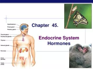
Chapter 45. Hormones and the Endocrine System. The Body’s Long-Distance Regulators Animal hormones Chemical signals secreted into blood and communicate regulatory messages within the body Hormones reach all parts of the body But only certain cells, target cells , respond.

E N D
Chapter 45 • Hormones and the Endocrine System
The Body’s Long-Distance Regulators • Animal hormones • Chemical signals secreted into blood and communicate regulatory messages within the body • Hormones reach all parts of the body • But only certain cells, target cells, respond
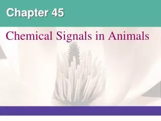
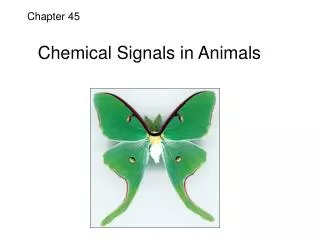
Insect metamorphosis • regulated by hormones Figure 45.1
The endocrine system and the nervous system act individually and together in regulating an animal’s physiology
nervous system • Conveys high-speed electrical signals along specialized cells called neurons • endocrine system • Secretes hormones that coordinate slower but longer-acting responses to stimuli
Overlap Between Endocrine and Nervous Regulation • Function together in maintaining homeostasis, development, and reproduction
Specialized nerve cells known as neurosecretory cells • Release neurohormones into the blood
Pathway Example Pathway Example Example Pathway Low blood glucose Hypothalamic neurohormone released in response to neural and hormonal signals Stimulus Stimulus Stimulus Suckling Receptor protein Sensory neuron Sensory neuron Pancreas secretes glucagon ( ) Hypothalamus/ posterior pituitary Hypothalamus Endocrine cell Neurosecretory cell Blood vessel Neurosecretory cell Hypothalamus secretes prolactin- releasing hormone ( ) Posterior pituitary secretes oxytocin ( ) Blood vessel Blood vessel Target effectors Liver Anterior pituitary secretes prolactin ( ) Target effectors Smooth muscle in breast Glycogenbreakdown,glucose releaseinto blood Response Endocrine cell Blood vessel (a) Simple endocrine pathway Response Milk release (b) Simple neurohormone pathway Target effectors Mammary glands Milk production Response Figure 45.2a–c (c) Simple neuroendocrine pathway Control Pathways and Feedback Loops • 3 types of hormonal control pathways
Common feature of control pathways feedback loop connecting the response to the initial stimulus e.g. Negative feedback
Hormones bind to target cell receptors pathways cell responses • Hormones bloodstream target cells
3 classes of hormones • Proteins and peptides • Amines derived from amino acids • Steroids
Signaling involves: • Reception • Signal transduction • Response
SECRETORY CELL Hormone molecule VIA BLOOD Signal receptor TARGET CELL Signal transduction pathway Cytoplasmic response OR DNA Nuclear response NUCLEUS Cell-Surface Receptors for Water-Soluble Hormones • Embedded in membrane, project from cell surface Figure 45.3a (a) Receptor in plasma membrane
Binding of a hormone transduction pathway specific responses
The same hormone may have different effects on target cells that have • Different receptors • Different signal transduction pathways • Different proteins for carrying out the response
Different receptors different cell responses Epinephrine Epinephrine Epinephrine a receptor b receptor b receptor Glycogendeposits Vessel dilates Vessel constricts Glycogen breaks down and glucose is released from cell (a) Intestinal blood vessel (b) Skeletal muscleblood vessel (c) Liver cell Different intracellular proteins different cell responses • e.g. epinephrine has multiple effects Figure 45.4a–c
Intracellular Receptors for Lipid-Soluble Hormones • Steroids Enter target cells, bind to protein receptors in the cytoplasm or nucleus
SECRETORY CELL Hormone molecule VIA BLOOD TARGET CELL Signal receptor DNA Signal transduction and response mRNA NUCLEUS Synthesis of specific proteins • Results in regulation of specific genes Figure 45.3b (b) Receptor in cell nucleus
Paracrine Signaling • Signals elicit responses in nearby target cells
e.g. Prostaglandins help regulate the aggregation of platelets in blood clots Figure 45.5
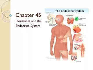
The hypothalamus and the pituitary gland • Control much of the endocrine system
Hypothalamus Pineal gland Pituitary gland Thyroid gland Parathyroid glands Adrenal glands Pancreas Ovary (female) Testis (male) • The major human endocrine glands Figure 45.6
Hypothalamus Neurosecretory cells of the hypothalamus Axon Posterior pituitary Anterior pituitary ADH Oxytocin HORMONE Mammary glands, uterine muscles Kidney tubules TARGET Figure 45.7 Hypothalamus and Pituitary Gland • Direct-acting hormonesare stored in and released from the posterior pituitary
Neurosecretory cells of the hypothalamus Tropic Effects Only FSH, follicle-stimulating hormone LH, luteinizing hormone TSH, thyroid-stimulating hormone ACTH, adrenocorticotropic hormone Nontropic Effects Only Prolactin MSH, melanocyte-stimulating hormone Endorphin Nontropic and Tropic Effects Growth hormone Portal vessels Endocrine cells of the anterior pituitary Hypothalamic releasing hormones (red dots) Pituitary hormones(blue dots) Endorphin FSH and LH TSH MSH ACTH Prolactin Growth hormone HORMONE Bones Melanocytes Liver Mammary glands Testes or ovaries Thyroid Pain receptors in the brain TARGET Adrenal cortex Figure 45.8 • Other hormonesare secreted into the blood and transported to the anterior pituitary
Antidiuretic hormone (ADH) • water reabsorption in the kidneys
Growth hormone (GH) • Promotes growth and has diverse metabolic effects
Thyroid Hormones • Produces 2 iodine-containing hormones, T3 & T4
Hypothalamus Anterior pituitary TSH Thyroid T3 T4 + • The hypothalamus and anterior pituitary • Control the thyroid through negativefeedback Figure 45.9
Thyroid hormones • Stimulate metabolism and influence development and maturation
Figure 45.10 • Hyperthyroidism, excessive secretion of thyroid hormones Graves’ disease in humans
Calcitonin Thyroid gland releases calcitonin. Reduces Ca2+ uptake in kidneys Stimulates Ca2+ deposition in bones Rising blood Ca2+ level Blood Ca2+ level declines to set point Homeostasis: Blood Ca2+ level (about 10 mg/100 mL) Falling blood Ca2+ level Blood Ca2+ level rises to set point Stimulates Ca2+ release from bones Parathyroid gland PTH Increases Ca2+ uptake in intestines Stimulates Ca2+ uptake in kidneys Active vitamin D Figure 45.11 Parathyroid Hormone and Calcitonin: Control of Blood Calcium • 2 antagonistic hormones, parathyroid hormone (PTH) and calcitonin
Insulin and Glucagon: Control of Blood Glucose • 2 types of cells in the pancreas secrete insulin and glucagon, antagonistic hormones that maintain glucose homeostasis and are found in the islets of Langerhans
Glucagon • produced by alpha cells • Insulin • produced by beta cells
Body cells take up more glucose. Insulin Beta cells of pancreas are stimulated to release insulin into the blood. Liver takes up glucose and stores it as glycogen. STIMULUS: Rising blood glucose level (for instance, after eating a carbohydrate- rich meal) Blood glucose level declines to set point; stimulus for insulin release diminishes. Homeostasis: Blood glucose level (about 90 mg/100 mL) STIMULUS: Dropping blood glucose level (for instance, after skipping a meal) Blood glucose level rises to set point; stimulus for glucagon release diminishes. Alpha cells of pancreas are stimulated to release glucagon into the blood. Liver breaks down glycogen and releases glucose into blood. Glucagon Figure 45.12 • Maintenance of glucose homeostasis
Insulin reduces blood glucose levels by • Cellular uptake of glucose • Slowing glycogen breakdown in the liver • Fat storage
Glucagon increases blood glucose levels by • Conversion of glycogen to glucose in the liver • Breakdown of fat and protein into glucose
Diabetes Mellitus • Perhaps the best-known endocrine disorder • Deficiency of insulin or a decreased response to insulin in target tissues Elevated blood glucose levels
Type I diabetes (insulin-dependent) • autoimmune disorder, immune system destroys the beta cells of the pancreas • Type II diabetes (non-insulin-dependent) • deficiency of insulin or, reduced responsiveness of target cells due to some change in insulin receptors
Adrenal Hormones: Response to Stress • adjacent to kidneys • adrenal medulla & adrenal cortex • secretes epinephrine and norepinephrine • Mediate various fight-or-flight responses
Glucocorticoids, e.g. cortisol • glucose metabolism • Mineralocorticoids, e.g. aldosterone • salt and water balance
Stress and the adrenal gland Stress Nerve signals Hypothalamus Spinal cord (cross section) Releasing hormone Nerve cell Anterior pituitary Blood vessel Adrenal medulla secretes epinephrine and norepinephrine. Nerve cell Adrenal cortex secretes mineralocorticoids and glucocorticoids. ACTH Adrenal gland Kidney (a) Short-term stress response (b) Long-term stress response Effects of epinephrine and norepinephrine: Effects of mineralocorticoids: Effects of glucocorticoids: 1. Glycogen broken down to glucose; increasedblood glucose 1. Retention of sodiumions and water bykidneys 1. Proteins and fatsbroken down andconverted to glucose,leading to increasedblood glucose 2. Increased blood pressure 3. Increased breathing rate 2. Increased bloodvolume and bloodpressure 4. Increased metabolic rate 5. Change in blood flow patterns, leading to increased alertness and decreased digestive and kidney activity 2. Immune system may be suppressed Figure 45.13a,b
Gonadal Sex Hormones • The gonads (testes and ovaries) • Produce androgens, estrogens, and progestins
Testes synthesize androgens (e.g. testosterone) • Development and maintenance of the male reproductive system, increase in muscle mass
Estrogen • maintenance of female reproductive system and the development of female secondary sex characteristics • Progesterone • preparing and maintaining the uterus
Melatonin and Biorhythms • Pineal gland (located within brain) • Secretes melatonin
Release of melatonin • Controlled by light/dark cycles • Function of melatonin biological rhythms associated with reproduction
Brain Neurosecretory cells in the brain produce brain hormone (BH), which is stored in the corpora cardiaca (singular, corpus cardiacum) until release. 1 Neurosecretory cells Brainhormone (BH) Corpus cardiacum Corpus allatum LowJH Prothoracicgland Juvenile hormone (JH), secreted by the corpora allata, determines the result of the molt. At relatively high concen- trations of JH, ecdysone-stimulated molting produces another larval stage. JH suppresses metamorphosis. But when levels of JH fall below a certain concentration, a pupa forms at the next ecdysone-induced molt. The adult insect emerges from the pupa. 4 Ecdysone Juvenilehormone(JH) BH signals its main target organ, the prothoracic gland, to produce the hormone ecdysone. 2 EARLYLARVA LATERLARVA PUPA ADULT Ecdysone secretion from the prothoracic gland is episodic, with each release stimulating a molt. 3 • e.g. insects • Molting and development controlled by hormones Figure 45.15