Download

1 / 72
720 likes | 757 Views
CLONING VECTORS. Dr. S. Rubanraj Dept of Mathematics St. Joseph’s College Trichy. Vectors.

E N D
CLONINGVECTORS Dr. S. Rubanraj Dept of Mathematics St. Joseph’s College Trichy
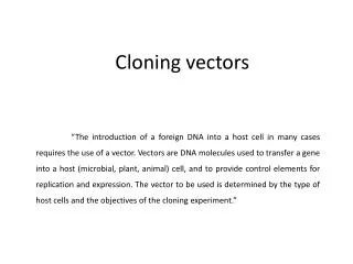
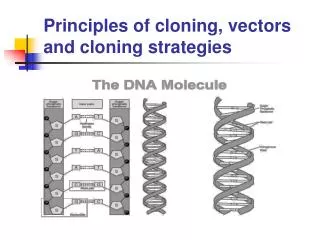
Vectors Vector is an agent that can carry a DNA fragment into a host cell in which it is capable of replication. If it is used only for reproducing the DNA fragment, it is called a cloning vector. If it is used for expression of foreign gene, it is called an expression vector. Vectors are ship for carrying the target DNA into a host cell. Cloning vector – used for obtaining millions of copies of cloned DNA segment.Used for creating genomic library or preparing the probes or genetic engineering experiments or other basic studies. Expression vector – allows expression of cloned gene, to give the product (protein). This can be achieved through the use of promoters and expression cassettes and regulatory genes. Used for transformation to generate trangenic plants, animals or microbes where cloned gene expresses to give the product.
Properties of a good vector: (1) It should be autonomously replicating i.e. it should have ori region. (2) It should contain at least one selectable marker e. g. gene for antibiotic resistance (tetR for tetracycline resistance). (3) It should have unique restriction enzyme site (only one site for one RE) for different REs (preferably in one of the marker genes) to insert foreign DNA. (4) It should be preferably small in size for easy handling. (5) It should have relaxed control of replication so that multiple copies can be obtained. (6) It should contain specific control systems like promoters, terminators, ribosome binding sites etc so that the cloned DNA should express properly.
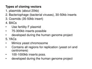
TYPES Vectors are of different types depending on the host. These are as follows: 1. Bacterial vectors 2. Yeast vectors 3. Plant vectors 4. Animal vectors
Bacterial vectors E.coli is the most commonly used bacterium for gene cloning though other bacteria such as Bacillus are also used. Vectors for cloning in these bacteria are described below: Vectors for cloning in E.coli A number of vectors are used for cloning in E.coli. Theses are categorized as plasmids, phages, cosmids, phagemids and bacterial artificial chromosomes.
PLASMIDS Plasmids are classified1. bytheirabilitytobetransferredtoother bacteria ConjugativeThesexual transfer of plasmidstoanotherbacteriumthrough a pilus. thoseplasmidspossessthe 25 genes requiredfor transfer. Non-conjugativeNon-conjugativeplasmidsdon’tinitiateconjugation. They can onlybetransferredwiththehelp of conjugativeplasmids. mobilisableAnintermediateclass of plasmids are mobilisable, and carryonly a subset of the genes requiredfor transfer. Theseplasmids can 'parasitise' anotherplasmid, transferring at highfrequency in thepresence of a conjugativeplasmid
2. byfunction 1. Fertility-(F) plasmids, They are capable of conjugation (theycontainsthe genes forthepili). Resistance-(R) plasmids, contain gene (s) that can buildresistanceagainstoneorseveralantibioticsorpoisons. Col-plasmids, contain genes codingforcolicines, proteinsthat can killother bacteria. Degradativeplasmids, abletodigestunusualsubstances, e.g., tolueneorsalicylicacid. Virulenceplasmids, turn a bacteriuminto a pathogen. addictionsystem. Theseplasmids produce both a long-livedpoison and a short-livedantidote. Daughtercellsthatretain a copy of theplasmidsurvive, while a daughtercellthatfails toinherittheplasmiddiesorsuffers a reducedgrowth-ratebecause of thelingeringpoisonfromtheparentcell.
Conjugativeplasmids The sexual transfer of plasmidstoanotherbacteriumthrough a pilus. Thoseplasmids, F plasmids, possessthe 25 genes requiredfor transfer. Plasmid forms: 1.covalently closed circles –both strands of DNA intact 2. open circles-only one of the two strand is intact 3. Linear. 4.Supercoiled circles.
Plasmid vectors • Plasmids are autonomously replicating circular, double stranded DNA molecules found in bacteria. • They have their own origin of replication (ori region), and can replicate independently of the host chromosome. • The size of plasmids ranges from a few kb to 200 kb. Plasmid vectors are often used for cloning DNA segments of small size (upto 10 kilobases). • Single copy plasmid - maintained as single copy per cell • Multicopy plasmid - maintained as 10-20 copies per cell • Plasmids under relaxed control of replication – over 1000 copies per cell – used as cloning vectors. • In each bacterial cell about 20-25 plasmids are maintained under normal growth condition. • Some of the commonly used plasmid vectors are described below:
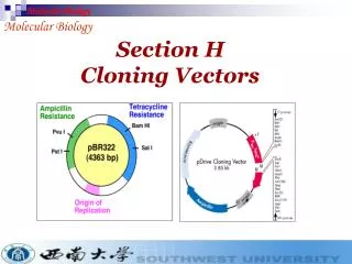
pBR322 • The first plasmid vector that has been constructed artificially is pBR322. • It is named after the scientists Bolivar and Rodriguiz who constructed it in 1977. • It is 4362bp in size and most widely used cloning vector. • It has an origin of replication derived from a colicin-resistance plasmid (ColE1). • This origin allows a fairly high copy number, about 100 copies of the plasmid per cell. • Plasmid pBR322 carries two selectable markers viz. genes for resistance to ampicillin (Apr) and tetracycline (Tcr).
Several (over 40 enzymes) unique RE sites are present within these genes for insertion of foreign DNA (Fig 1). • EcoRIV, BamHI,SphI, SalI,XmaIII, and NruI are present within the gene coding for tetracycline resistance, two sites (HindIII and ClaI) within the promoter of the tetracycline resistance gene and the three sites (PstI, PvuI and ScaI) within the βlactamase gene that provide resistance to ampicillin • When a foreign DNA segment is inserted in any of these genes, the antibiotic resistance by that particular gene is lost. This is called insertional inactivation. • For instance, insertion of a restriction fragment in the SalI site of the Tcrgene inactivates that gene. • One can still select for Apr colonies, and then screen to see which ones have lost Tcr.
Figure pBR322 The map shows the positions of the ampicillin-resistance gene (ampR), the tetracycline-resistance gene (tetR), the origin of replication (ori) and the recognition sequences for seven restriction endonucleases.
The plasmid pBR322 is one of the most commonly used E.coli cloning vectors. pBR322 is 4361 bp in length and contains: (1) the repliconrep responsible for the replication of plasmid (source – plasmid pMB1); (2) rop gene coding for the Rop protein, which promotes conversion of the unstable RNA I – RNA II complex to a stable complex and serves to decrease copy number (source – plasmid pMB1); (3)bla gene, coding for beta-lactamase that confers resistance to ampicillin (source – transposon Tn3); (4) tet gene, encoding tetracycline resistance protein (source – plasmid pSC101).
pUC • A series of small plasmids (about 2.7 kb) have been developed (by Messings and co-workers in1983) at the University of California and hence the name pUC e.g. pUC7, 8,9,12,13, 18 and 19 etc. • These are high copy number plasmids that carry an ampicillin resistance gene and an origin of replication, both from pBR322. • They also have a multiple cloning site (MCS) – a sequence of DNA that carries unique sites for many REs. • The MCS contains a portion of lacZ gene that codes for the enzyme β-galactosidase. • When such plasmids are introduced into E. coli, the colonies are blue on plates containing X-gal (5-bromo-4-chloro-3-indolyl-β-d-galactopyranoside, the substrate for β- galactosidase) and IPTG (isopropyl thiogalactoside, an inducer).
Recombinants and non-recombinants can therefore be distinguished simply by plating the transformed cells onto agar containing ampicillin and X-gal. • All colonies that grow on this medium are made up of transformed cells because only transformants are ampicillin resistant. • Blue colonies contain cells with functional β-galactosidase enzymes and hence with undisrupted lacZ′ genes these colonies are therefore non-recombinants. • The white colonies comprise cells without β-galactosidase activity and hence with disrupted lacZ′ genes; these are recombinants. • Thus cells containing recombinant plasmids form white (not blue) colonies.
Must have: 1) Ori 2) A dominant selectable Marker 3) Cleavage sites for cloning 4) (high copy no.) The plasmid cloning vector pUC19. This plasmid has an origin of replication (ori), an ampR selectable marker, and a polylinker located within part of the -galactosidase gene lacZ+.
Phage vectors • Bacteriophages or phages are viruses that specifically infect bacteria. • The phage particle attaches to the outer surface of bacterium and injects its DNA into the cell. • The phage DNA is then replicated inside the host and its genes are expressed to make phage capsid proteins and new phage particles are assembled and released from the bacterium. • Phage vectors can accommodate more DNA (upto 25 kb) than plasmids and are often used for preparation of genomic libraries. • They also have higher transformation efficiency as compared to plasmids.
The main reason for seeking a different type of vector was the inability of plasmids such as pBR322 and pUC8 to handle DNA fragments greater than about 10 kb in size, larger inserts undergoing rearrangements or interfering with the plasmid replication system in such a way that the recombinant DNA molecules become lost from the host cells. • The first attempts to develop vectors able to handle larger fragments of DNA centered on bacteriophage λ. • Two bacteriophages namely, Lambda (λ) and M13 have been commonly used for construction of vectors for cloning in E. coli. • The phage can have two modes of life cycles i.e. lytic and lysogenic. • During lytic cycle, it replicates independently in the host cell and produces a large number of phage particles which are released by lysis of the host. Alternatively, it can take up lysogenic growth, meaning that it integrates its DNA into the bacterial chromosome and multiplies along with it.
(N, cro, cI genes) Map of the λ chromosome of wild type . The genes are not essential for phage growth and can be deleted or replaced without seriously impairing the infectious growth cycle
The lysogenic infection cycle of bacteriophage λ The special feature of the lysogenic cycle is the insertion of the phage genome into the bacterium's chromosomal DNA, where it can remain quiescent for many generations.
Three temporal stages of λ transcription occurs in the lytic cycle : • early gene transcription establishes the lytic cycle (in competition with lysogeny) - early transcription proceeds from promoters PL and PR, stop at termination sites tL and tR1 -transcription is subject to repression by the product of the cI gene . • middle gene products replicate and recombine the DNA – It is directed by N gene product - PL and PR transcripts extend into genes such as red, O and P necessary for the middle stage. • late gene products are packaged into mature phage particles - The cro product accumulate and prevents transcription from PL and PR. Q product is responsible for middle-to-late switch and the Q product specifically anti-terminates the short PR ´ transcript - many mature phage particles are produced.
Lambda (λ) phage vectors • Lambda is a temperate bacteriophage with a genome size of 48.5 kb. Its entire DNA sequence is known. • The lambda genome is a linear, double-stranded molecule with single-stranded, complementary ends. These ends can hybridize with each other (and do so when the DNA is within an infected cell) and are thus termed cohesive (cos) sites. • The λ genome is 48.5 kb, of which some 15 kb or so is ‘optional’ in that it contains genes that are only needed for integration of the phage DNA into the E. coli chromosome. These segments can therefore be deleted without impairing the ability of the phage to infect bacteria and direct synthesis of new λ particles by the lytic cycle.
Two types of vector have been developed: • Insertion vectors, in which part or all of the optional DNA has been removed and a unique restriction site introduced at some position within the trimmed down genome; • Replacement vectors, in which the optional DNA is contained within a stuffer fragment, flanked by a pair of restriction sites, that is replaced when the DNA to be cloned is ligated into the vector. • The λ genome is linear, but the two natural ends of the molecule have 12-nucleotide single-stranded overhangs, called cossites, which have complementary sequences and so can base-pair to one another.
Insertional vectors have one unique restriction site for a particular restriction enzyme and can accommodate 6-7 kb DNA. • Examples of insertional vectors are λgt10, λgt11 and λZAP II. • On the other hand, replacement vectors have two cleavage sites for a restriction enzyme and can accommodate up to 25 kb DNA. • When vector is cut with a restriction endonuclease, a stuffer fragment is removed and replaced with a foreign DNA. • Some examples of replacement vectors are EMBL3, EMBL3A, EMBL4, λDASH, λFIX, GEM11 and GEM12.
Cloning vectors based on bacteriophage λ In the λ genome, the genes are arranged into functional groups. For example, the region marked as ‘protein coat’ comprises 21 genes coding for proteins that are either components of the phage capsid or are required for capsid assembly, and ‘cell lysis’ comprises four genes involved in lysis of the bacterium at the end of the lytic phase of the infection cycle. The regions of the genome that can be deleted without impairing the ability of the phage to follow the lytic cycle are indicated in green. (B) The differences between a λ insertion vector and a λ replacement vector.
Bacteriophage lambda can be reconstituted in a test tube by simply mixing phage DNA with a mixture of phage proteins, an infective viral particle with the DNA inside the phage head can be produced. This process is called in vitro packaging. • There is a strict size requirement for the piece of DNA that goes into the phage head. That is, it should not be more than 52 kb and less than 38 kb. • This feature allows only the recombinants to be packaged inside the phage head. • In addition, some lambda phage vectors have a stuffer fragment that carries the β-galactosidase gene. • When it is removed or when foreign DNA is cloned within the gene, β-galactosidase activity may be abolished. The accompanying loss of activity may be used to select recombinant clones.
Cloning with a λ insertion vector The linear form of the vector is shown at the top of the diagram. Treatment with the appropriate restriction endonuclease produces the left and right arms, both of which have one blunt end and one end with the 12-nucleotide overhang of the cos site. The DNA to be cloned is blunt ended and so is inserted between the two arms during the ligation step. These arms also ligate to one another via their cos sites, forming a concatamer. Some parts of the concatamer comprise left arm-insert DNA-right arm and, assuming this combination is 37–52 kb in length, will be enclosed inside the capsid by the in vitro packaging mix. Parts of the concatamer made up of left arm ligated directly to right arm, without new DNA, are too short to be packaged.
Bacteriophage infection is visualized as a plaque on a lawn of bacteria
DNA cloning with single stranded DNA vectors • These coliphages are developed as cloning vectors • This phage particles have dimensions 900 nm × 9 nm contain a single-stranded circular DNA molecule (6407 (M13) or 6408 (fd) nucleotides long). • they have many advantages over other vectors • M13, f1 and fd are filamentous coliphagescontaining a circular single-stranded DNA molecule.
The biology of the filamentous coliphages • The phages only infect enteric bacteria harbouring F pili • the end of the F pilus is the adsorption site . • infected cells continue to grow and divide, and extrude virus particles. • Up to 100 phage particles may be released into the medium per cell per generation • Replication of phage DNA does not result in host-cell lysis. • The process of phage transfect and release – • 1. The single-stranded phage DNA enters the cell • 2. the single-stranded phage DNA is released.
The single-stranded phage DNA enters the cell • the virus discard the capsid proteins • the viral DNA passes into the cell • converted to a double-stranded replicative form . • the decapsidation and replication are tightly coupled. The RF multiplies rapidly inside the cell until about 100 RF molecules.
a viral-encoded protein accumulated. • The protein binds to the viral strand and prevents synthesis of the complementary strand. • Replication of the RF becomes asymmetric • only viral single strands are synthesized.
Why use single-stranded vectors? • Sequencing by dideoxy method required single-stranded DNA • oligonucleotide-directed mutagenesis required single-stranded DNA. • certain probe preparation required single-stranded DNA. • In summary filamentous phages possess all the advantages of plasmids and producing particles containing single-stranded DNA in an easily obtainable form.
M13 Phage vectors • M13 is a filamentous bacteriophage of E. coli and contains a single stranded circular DNA of 7.2 kb. • A series of vectors (M13 mp series) have been developed from this phage. • These vectors have a polylinker with unique restriction enzyme sites in lac Z gene that complements host (e.g. JM 103 or JM 104). • Screening of recombinants is done based on formation of blue/white plaques. • M13 vectors are used for obtaining sufficient quantity of DNA for sequencing by Sanger's dideoxy chain termination method.
A typical cosmid pJB8 is 5.4 kb in size and carries the ampicillin-resistance gene (ampR), a segment of λ DNA containing the cos site, and an Escherichia coli origin of replication (ori).
cosmid vector The cosmid vector is a combination of the plasmid and bacteriophage lambda. It is small (5-7 kb) circular DNA containing an origin for DNA replication (ori), selectable markers and restriction sites from plasmid plus a sequence from lambda needed for packaging the DNA (cos site). Cosmids may be used to clone large DNA molecules of up to 45 kb. They also have high transformation efficiency. Some examples of cosmid vectors include pJB, PWE and SuperCos series.
Vectors for cloning in yeast The discovery of a 2μm plasmid in most strains of Saccharomycescerevisiae led to the development of cloning vectors in yeast. The 2μm plasmid is 6 kb in size. It is present in 50-100 copies per cell. A number of shuttle vectors based on 2μm plasmid and bacterial plasmids have been constructed which can replicate either in E.coli or yeast. Yeast plasmid vectors are of four types, yeast episomal plasmids (YEps), yeast integrative plasmids (YIps) yeast replicative plasmids (YRps) and yeast centromeric plasmids (Ycps). In addition to plasmid vectors, yeast artificial chromosomes (YACs) are also used as vectors for cloning large pieces of DNA.
i) Yeast episomal plasmids (YEps) These are derived from 2μm plasmid. Some YEps contain the entire 2μm plasmid; others include just the 2μm origin of replication. An example of latter type is YEp13. It is a shuttle vector and can be replicated both in E.coli and yeast. It contains 2μm origin of replication, yeast gene leu2 as selectable marker and entire sequence of pBR322.The leu2 gene codes for an enzyme involved in biosynthesis of amino acid leucine. YEps may replicate autonomously or integrate in one of the yeast chromosomes by homologous recombination. They have high transformation frequency of 10,000 to 100,000 transformants/ μg DNA.
ii) Yeast integrative plasmids (YIps) These are basically bacterial plasmids carrying a yeast gene. YIp5 is an example of yeast integrative plasmid. It has ura3 gene inserted in pBR322. The gene ura3 codes for an enzyme involved in biosynthesis of pyrimidine nucleotides and acts as selectable marker. The plasmid cannot replicate autonomously as it lacks 2μm origin of replication and survives by integrating in yeast chromosomal DNA. They have very low transformation frequency, less than 100 transformants/ μg DNA.
iii) Yeast replicative plasmids (YRps) They carry a part of chromosomal DNA with an origin of replication and one or two selectable markers and are capable of independent replication. They have transformation frequency between 1000 and 10,000 transformants/ μg DNA. iv) Yeast centromeric plasmids (YCps) These are shuttle vectors that behave as small chromosomes and replicate only once during each cell divison. They contain i) origin of replication called ARS sequence , ii) CEN sequence (for proper segregation of chromosomes) and iii) a selectable marker such as leu2 from yeast and sequences from bacterial plasmid having ori region and selectable marker (Apr). They are stably maintained at one copy per cell.
v) Yeast Artificial Chromosomes (YACs) YACs are artificial chromosomes that replicate in yeast cells. Main features of these vectors are: 1. Autonomously replicating sequence (ARS) necessary for the replication in yeast cells (Fig 6). 2. Telomeres (TEL), which are ends of chromosomes involved in the replication and stability of chromosomes. 3. A yeast centromere (CEN), required for proper segregation of chromosomes 4. Selectable markers that allow the easy isolation of yeast cells that have taken up the artificial chromosome. 5. Unique RE sites. YACs are capable of carrying a large DNA fragment (up to 3000 kb), but their transformation efficiency is very low.