Download
1 / 18
260 likes | 554 Views
Prenatal Testing. Introduction. Tests are available during pregnancy to check the health of a baby. What conditions can be found? Down syndrome. Neural tube defects such as spina bifida and anencephaly. Birth defects such as congenital heart

E N D
Introduction • Tests are available during pregnancy to check the health of a baby. • What conditions can be found? • Down syndrome. • Neural tube defects such as spina bifida and • anencephaly. • Birth defects such as congenital heart • conditions, malformed kidneys, missing limbs, • trisomy 13 and trisomy 18.
Neural Tube Defects • A baby’s brain and spine develop from a neural tube in the first four weeks of pregnancy. A neural tube defect occurs when the tube does not fully develop. Spina bifida occurs when the tube does not completely close along the spine. Other neural tube defects include anencephaly and encephalocele in which the brain and skull do not develop properly. About one in every 700 pregnancies is affected by a neural tube defect. This defect often causes the baby to be stillborn or die shortly after birth.
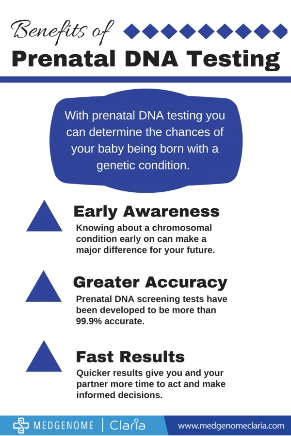
Down Syndrome • Down syndrome • Down syndrome is a condition that results in a range of physical and intellectual disabilities. It is caused by an extra copy of chromosome 21 in every cell (trisomy 21). Down syndrome occurs in about one in 400 pregnancies. Women of any age can have a Down syndrome pregnancy, however this risk is higher for women over 35 years (see FAQ section). Other types of chromosome conditions are trisomy 13 and trisomy 18.
Amniocentesis • Amniocentesis is a medical procedure performed on a pregnant woman to withdraw a small amount of amniotic fluid from the sac surrounding the foetus. By about the 16th week of pregnancy, the developing baby is suspended in around 130ml of amniotic fluid, which the baby constantly swallows and excretes. The goal of amniocentesis is to examine a tiny amount of this fluid to obtain information about the baby - including its sex - and to detect physical abnormalities such as Down's syndrome or spina bifida. Amniocentesis is only performed on women thought to be at higher risk of delivering a child with a birth defect.
Problems detected by Amniocentesis Amniocentesis can detect a number of disorders that will affect babies, while they are still a small foetus in the uterus. These conditions include: • Down's syndrome. • Neural tube defects, such as spina bifida. • Cystic fibrosis. • Genetic disorders - amniotic fluid samples can be DNA tested to identify a wide range of genetic disorders, including Fragile X syndrome, phenylketonuria, Tay-Sachs disease and sickle cell disease.
Amniocentesis – The Procedure Before having amniocentesis, it is usual for the woman and her partner to be counselled on the risks of the procedure. Amniocentesis is performed between 16 and 20 weeks into the pregnancy. The woman lies down, and the position of the foetus and the placenta are determined by an ultrasound scan. When the doctor is sure of a safe spot, they swab the woman's belly with antiseptic and inject a local anaesthetic into the skin. Using a long, thin needle, the doctor extracts about 15 to 20ml (approximately three teaspoons) of amniotic fluid. This takes about 30 seconds or so. The foetus is checked afterwards to make sure all is well. The entire procedure can take around 90 minutes.
Ultrasounds Ultrasound is the use of high frequency sound waves to make an image. When used in pregnancy, an ultrasound gives a picture of the developing baby in the uterus (womb). Ultrasound is often considered part of the routine care for all pregnant women.
Timing • Current knowledge suggests that there is no harmful effect on the mother or the developing baby. Ultrasound does not involve X-rays. Ultrasound scanning can take place at any time during a pregnancy. • For testing purposes there are two ideal times for doing an ultrasound scan. • 1st trimester ultrasound also called nuchal translucency ultrasound is done at 11-13* weeks of pregnancy. This scan measures the skin thickness at the back of the baby's neck (nuchal translucency). When this measurement is increased it can indicate an increased risk of having a baby with Down syndrome or another problem. When an ultrasound is performed at an accredited centre the result can be combined with the first trimester blood test. • If this test indicates an increased risk the woman will be offered diagnostic testing, either CVS or amniocentesis. • 2nd trimester ultrasound sometimes called the fetal anomaly scan is done at 18-20 weeks of pregnancy. This scan is used to identify structural problems such as limb or heart defects, in the baby. It does not detect all abnormalities. • If problems are detected the woman will be offered further testing.
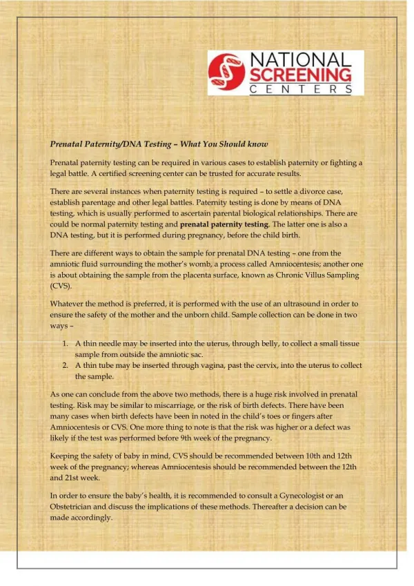
Chronic Villus Sampling • Chorionic villus sampling (CVS) is a very accurate antenatal test that detects chromosomal abnormalities such as Down syndrome. It takes placental tissue, as opposed to baby cells used in an amniocentesis. Its main advantage over amniocentesis is that it can be performed earlier -- between the 10th and 14th weeks of pregnancy. You generally can't have an amniocentesis until 15 weeks of pregnancy. Some women choose CVS because they already have a child with a birth defect, or they have a family history of a genetic disease such as Tay Sachs and they want to know as soon as possible whether this baby is affected. Many hospitals offer CVS and it has become a popular alternative to amniocentesis.
The Procedure • You'll be asked to lie down for the test. First you have an ultrasound scan to confirm how many weeks pregnant you are, and to locate the placenta. The ultrasound examination might be uncomfortable when the doctor presses down on your tummy with the transducer. However, you'll be able to see your baby on the screen which is a bonus! Your doctor will pass a needle thorugh your abdomen to reach the placenta. In a very few cases you may be recommended to have a transvaginal CVS where a fine tube will be thread through your vagina. For a transabdominal CVS, you will be given a local anaesthetic to numb the wall of your abdomen before the needle is inserted. Your doctor will then extract a fragment of chorionic villi -- tiny fingerlike projections on the placenta. The cells taken from the placenta are full of genetic information that can be analysed to reveal the chromosomal make-up and the sex of your baby.
Foetal Heart Monitoring Foetal monitoring is the recording of the baby's heart rate and the mother's contractions during labour. Devices are connected to the mother's abdomen and to the baby. This is done in two ways: External monitoring uses external belts around the mother's abdomen. • Internal monitoring involves placing a monitor electrode on the baby's scalp. A thin tube or catheter is also inserted into the uterus via the vagina to monitor contractions.
Candidates of Foetal Heart Monitoring • The contractions of the uterus during labour decrease the amount of blood flowing to the placenta, the organ that normally attaches to the uterus, connecting the developing foetus to the mother and supplies nutrition and oxygen to the foetus. Contractions also decrease the blood flow to the foetal umbilical cord, which inserts into the developing baby's belly button and connects the foetus to the placenta. The decreased blood flow cuts down on the amount of oxygen getting to the baby. Labour and delivery can be risky to the foetus under normal conditions, but presents even more risk if the placenta is not functioning fully. In most hospital settings, the majority of women in labour undergo foetal monitoring to ensure a good outcome.
Conditions requiring FHM • diabetes in the mother • intrauterine growth retardation, a condition in which the foetus is not growing at an appropriate rate • past due pregnancy, of more than 42 weeks • preeclampsia, a toxic condition of pregnancy that may cause increased blood pressure, excessive swelling in the arms or legs, abnormal kidney function and disturbances in vision • eclampsia, a condition in which seizures develop in a woman with preeclampsia • chronic hypertension in the mother • multiple foetuses, such as twins or triplets • use of epidural anaesthesia, a type of anaesthesia in which the analgesia is given directly into the compartment that contains the spinal cord • use of drugs given to cause labour and delivery. These include oxytocin and prostaglandins. • suspected foetal distress • presence of green amniotic fluid due to meconium, or foetal bowel movement, which may cause meconium aspiration syndrome.
Blood Tests • Blood tests from the mother called "maternal serum screening" will measure the level of particular hormones and indicate the risk of a woman having a baby with Down syndrome or another problem. • Blood tests do not diagnose these conditions, but indicate whether or not there is an increased or decreased risk in this pregnancy. • There are two different types of maternal serum screening tests. You can only have one or the other, not both. • 1st trimester combined screening is a blood test done at 9-12 weeks of pregnancy (best during 10th week), combined with a nuchal translucency ultrasound scan done at 11-13* weeks of pregnancy. • This test will indicate the risk of having a baby with Down syndrome and another chromosome condition called Trisomy 18. As this is a combined test, a risk cannot be calculated using the blood sample alone. • If this test indicates an increased risk the woman will be offered a diagnostic test, either CVS or amniocentesis (see below). • 2nd trimester maternal serum screening also called the quadruple test is a blood test done at 14-20 weeks of pregnancy (best during 15th week). This test will indicate the risk of having a baby with Down syndrome, Trisomy 18 or neural tube defects (ie. spina bifida or anencephaly). • If this test indicates an increased risk for Down syndrome or Trisomy 18 the woman will be offered amniocentesis. If this test indicates an increased risk for spina bifida or anencephaly the woman will be offered an ultrasound examination to look for neural tube defects.