Download
1 / 68
820 likes | 1.46k Views
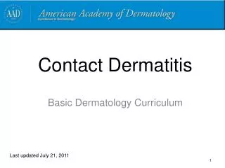
Contact Dermatitis. Medical Student Core Curriculum In Dermatology. Last updated July 21, 2011. Module Instructions.

E N D
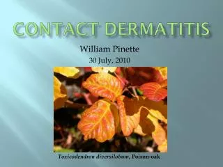
Contact Dermatitis Medical Student Core Curriculum In Dermatology Last updated July 21, 2011
Module Instructions • The following module contains a number of blue, underlined terms which are hyperlinked to the dermatology glossary, an illustrated interactive guide to clinical dermatology and dermatopathology. • We encourage the learner to read all the hyperlinked information.
Goals and Objectives • The purpose of this module is to help medical students develop a clinical approach to the evaluation and initial management of patients presenting with contact dermatitis. • By completing this module, the learner will be able to: • Identify and describe the morphology for contact dermatitis • Distinguish allergic contact dermatitis from irritant contact dermatitis • Recommend an initial treatment plan for a patient with allergic or irritant contact dermatitis • Determine when to refer a patient with contact dermatitis to a dermatologist
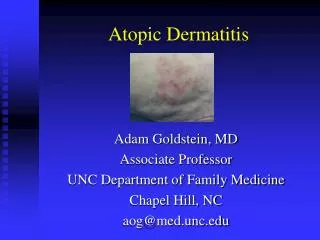
Dermatitis in General • Dermatitis or eczema is a pattern of cutaneous inflammation that presents with erythema, vesiculation, and pruritus in its acute phase • The chronic phase is characterized by dryness, scaling, lichenification, fissuring, and pruritus • There are multiple types of dermatitis: • seborrheic, atopic, dyshidrotic, nummular • This module will focus on contact dermatitis
Contact Dermatitis • Contact dermatitis is a skin condition created by a reaction to an externally applied substance • There are two types of contact dermatitis: • Irritant Contact Dermatitis (ICD) • Allergic Contact Dermatitis (ACD)
Case One Dr. Gary Richardson
Case One: History • HPI: Dr. Richardson is a 43-year-old neonatologist who presents with 3 days of intense itching and blisters on his neck, arms and legs. He noticed the eruption 2 days after a hike. Clobetasol ointment and oral diphenhydramine have been ineffective in controlling his symptoms. • PMH: none • Allergies: none • Medications: topical steroid, diphenhydramine • Family history: noncontributory • Social history: neonatologist, married, has a daughter • ROS: difficulty sleeping due to itching
Case One, Question 1 • Dr. Richardson’s exam shows erythematous plaques, consisting of confluent papules and weeping vesicles on his arms, legs, and neck bilaterally. Some of them are linear. What is the most likely diagnosis? • Allergic contact dermatitis • Bullous insect bites • Cellulitis • Herpes zoster • Urticaria
Case One, Question 1 Answer: a • Dr. Richardson’s exam shows erythematous papules and extensive weeping vesicles on his arms, legs, and neck bilaterally. Some of them are linear. What is the most likely diagnosis? • Allergic contact dermatitis • Bullous insect bites (usually scattered, not linear or grouped, no history of multiple bites) • Cellulitis (presents as a spreading erythematous, non-fluctuant tender plaque, often with fever) • Herpes Zoster (presents as a painful eruption of grouped vesicles in a dermatomal distribution) • Urticaria (presents as edematous plaques, not vesicles. The early lesions of allergic contact dermatitis could be mistaken for urticaria)
Allergic Contact Dermatitis • ACD occurs when contact with a particular substance elicits a delayed hypersensitivity reaction • The sensitization process requires 10-14 days • Upon re-exposure, dermatitis appears within 12-48 hrs • The most common cause is Rhus dermatitis, from poison ivy, poison oak, or poison sumac (all contain the resin – urushiol) • Other common causes include: • Fragrances • Formaldehyde • Preservatives • Topical antibiotics • Benzocaine • Vitamin E • Rubber compounds • Nickel
ACD: Clinical Findings • The main symptom of ACD ispruritus (itching) • Presents as eczematous, scaly edematous plaques with vesiculation distributed in areas of exposure • ACD is bilateral if the exposure is bilateral (e.g., shoes, gloves, ingredients in creams, etc.)
Back to Case One Dr. Richardson was diagnosed with Rhus allergic contact dermatitis
Poison Oak & Poison Ivy“Leaves of three- let them be” • Poison ivy leaves usually: • Are 3-15cm in length • Notched edges • Groups of 3s • Grows on hairy-stemmed vines or low shrubs • Turn colors in autumn • Poison oak leaves usually: • Are 3-7cm in length • Lobulated notched edges • Groups of 3, 5, or 7 • Grows on bush-like plants • Turn colors in autumn
Rhus Allergy • The initial episode occurs 7-10 days after exposure • On subsequent outbreaks the rash may appear within hours of exposure and usually within 2 days • Individual sensitivity is variable so the eruption may be mild to severe • Rhus dermatitis lasts from 10-21 days depending on the severity • Initial episode is the longest (up to 6 weeks!)
Rhus Allergy • Lesions begin as erythematous macules that become papules or plaques • Blisters often form over one to two days
Rhus Dermatitis • Linear streaks aid in diagnosis (from the linear contact of the plant) • Fomites can be contaminated by the plant oil and lead to recurrent eruptions
Case One, Question 2 • Dr. Richardson can’t sleep due to itching and has had no improvement with clobetasol ointment the past three days. What treatment do you recommend? • Oral cephalexin • 1% hydrocortisone lotion • Silver sulfadiazine cream • Six days of methylprednisolone (Medrol dose pack) • Two-week taper of oral prednisone
Case One, Question 2 Answer: e • Dr. Richardson can’t sleep due to itching and has had no improvement with clobetasol ointment the past three days. What treatment do you recommend? • Oral cephalexin (for gram positive bacterial infections) • 1% hydrocortisone lotion (not strong enough) • Silver sulfadiazine cream (for burns) • Six days of methylprednisolone (Medrol dose pack) (will likely get worse rebound after withdrawal) • Two-week taper of oral prednisone
Rhus Dermatitis Treatment • Most patients need minor supportive care • Topical steroids for localized involvement • Topical or oral antihistamines may improve pruritus • Oatmeal soaks/calamine lotion may soothe weeping erosions • Severe involvement may require oral steroids • In cases of failing potent topical steroids, or widespread • If given for less than 2-3 weeks, patients may relapse • Do not give short bursts of steroids for this reason
Rhus Allergy Prevention • Avoid the plants • Wash clothing, shoes, and objects after exposure (within 10 minutes if possible) • Apply barrier: clothing, OTC products which bind resin more than skin
Barbara Myers Case Two
Case Two: History • HPI: Barbara Myers is a 32-year-old woman who presents to the dermatology clinic with three months of severe itching, redness, and scaling on her eyelids. She has tried aloe vera and tea tree oil products, but they haven’t helped. • PMH: none • Allergies: shellfish • Medications: birth control pills • Family history: noncontributory • Social history: single; works as a bank teller • ROS: negative
Case Two: Skin Exam • On further questioning, Ms. Myers recently changed her eye shadow and moisturizer.
Case Two, Question 1 • Ms. Myers has bilaterally-symmetric, pruritic, erythematous, scaly, slightly lichenified plaques on her eyelids. What is the most likely diagnosis? • Allergic contact dermatitis • Atopic dermatitis • Rosacea • Seborrheic dermatitis
Case Two, Question 1 Answer: a • Ms. Myers has bilaterally-symmetric, pruritic, erythematous, scaly, slightly lichenified plaques on her eyelids. What is the most likely diagnosis? • Allergic contact dermatitis • Atopic dermatitis (does commonly involve the eyelid in adults and can be difficult to distinguish from allergic contact dermatitis) • Rosacea (would have papules and pustules, usually not itchy) • Seborrheic dermatitis (affects lid margin and eyebrow, but not eyelid, usually not itchy)
Eyelid Allergic Contact Dermatitis • Intensely pruritic • Scaling red plaques on upper > lower eyelids • Allergic contact dermatitis of the eyelid is often caused by transfer from the hands • Common causes: • Nail adhesive/polish • Fragrances and preservatives in cosmetics • Nickel
Evaluation of Dermatitis • Important to take a comprehensive history • Complete dermatologic assessment of the patient • The shape, configuration, and location of the dermatitis are useful clues in identifying the culprit allergen • Elimination of a suspected trigger may be both diagnostic and therapeutic • In chronic cases, patch testing is necessary to identify specific allergens
History Taking • In addition to the dermatitis-specific history (e.g., onset, location, temporal associations, treatment), be sure to ask about: • Daily skin care routine • All topical products • Occupation/hobbies • Regular and occasional exposures (e.g. lawn care products, animal shampoos)
Case Two, Question 2 • Ms. Myers has an allergic contact dermatitis, likely to her new eye shadow. What treatment would you recommend other than avoidance? • Clobetasol ointment • Desonide cream • Fluocinonide gel • Ketoconazole cream
Case Two, Question 2 Answer: b • Ms. Myers has an allergic contact dermatitis, likely to her new eye shadow. What treatment would you recommend other than avoidance? • Clobetasol ointment (too potent, class 1) • Desonide cream (for a limited period: twice daily for 1 week, followed by once daily for 1-2 weeks, then discontinue) • Fluocinonide gel (too potent, gels have alcohol and may burn on the eyelid, class 2) • Ketoconazole cream (treats fungal infection)
Steroid Potency • Regular use of Class 1, 2, or 3 steroids on thin skin will lead to steroid atrophy (thinning and easy bruising/purpura) • Also hypopigmentation in darker skin types • For the face: Class 6, 7 steroids (e.g., desonide) can safely be used intermittently during flares • If topical steroids are to be used on the eyelid for a period of more than one month, refer to an ophthalmologist for monitoring of intraocular pressure and the development of cataracts
Case Two, Question 3 • Ms. Myers has an allergic contact dermatitis that responds to topical steroids. What is the best test to confirm the cause of her rash? • Indirect immunofluorescent antibody (IIF) test • Patch testing • Prick skin testing • Radioallergosorbent test (RAST)
Case Two, Question 3 Answer: b • Ms. Myers has an allergic contact dermatitis that responds to topical steroids. What is the best test to confirm the cause of her rash? • Indirect immunofluorescent antibody (IIF) test (used for the diagnosis of antibody-mediated diseases, not contact dermatitis) • Patch testing • Prick skin testing (does not detect cell-mediated allergy) • Radioallergosorbent test (RAST) (used to detect type 1 hypersensitivity, not cell-mediated immunity)
Patch Testing • Patch testing is used to determine which allergens a patient with allergic contact dermatitis reacts against • A series of allergens are applied to the back, and they are removed after 2 days • On day 4 or 5, the patient returns for the results • Positive reactions show erythema and papules or vesicles • Identification of specific allergens helps the patient find products free of those allergens
Patch Testing • Example of a patient with patches (allergens) placed on the back
Identifying Allergens • Not all patients with ACD need patch testing • Refer patients when the allergen is unclear or the dermatitis is chronic • A positive reaction on patch testing does not mean that the patient’s rash is due to that specific allergen • Elimination of the rash with removal of the allergen confirms the clinical relevance of the positive patch test
Positive Patch Test • Positive patch test reactions at 96 hour reading • This patient had three positive reactions • Nickel, Balsam of Peru, and Fragrance • Avoidance of these allergens should improve their rash
ACD Treatment Avoid exposure to the offending substance
ACD Treatment • Treatment of the acute phase depends on the severity of the dermatitis • In mild to moderate cases, topical steroids of medium to strong potency for a limited course is successful • A short course of systemic steroids may be required for acute flares • Oatmeal baths or soothing lotions can provide further relief in mild cases • Wet dressings are helpful when there is extensive oozing and crusting • Chronic cases or patients with dermatitis involving over 10% of the BSA should be referred to a dermatologist
Patient calls 9 days after you performed a skin biopsy, reporting itching at the site
This 11-year-old girl presents with 3 months of an itchy rash on the sides of her nose and ears
Another Example of Nickel Dermatitis • Erythematous plaque with scattered papules above the umbilicus • Nickel dermatitis is the 2nd most common allergic contact dermatitis next to Rhus dermatitis
This respiratory therapist has an intermittent rash that clears when she goes on vacation
Latex Allergy • Latex allergy may present as a delayed or immediate hypersensitivity • Delayed hypersensitivity: • Patients develop an allergic contact dermatitis • Often presents on the dorsal surface of the hands • Immediate hypersensitivity: • May present with immediate symptoms such as burning, stinging, or itching with or without localized urticaria on contact with latex proteins • May include disseminated urticaria, allergic rhinitis, and/or anaphylaxis