Download
1 / 53
530 likes | 539 Views
Management of Difficult Colonic Lesions. Joint Hospital Surgical Grandround 10/2009. Challenge to Endoscopist. Lesions larger than 2cm are considered large colonic lesions Prevalence 0.8% - 5.2%. Challenge to Endoscopist. Polyps at difficult position Flexure Behind the folds

E N D
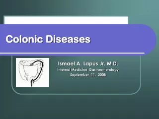
Management of Difficult Colonic Lesions Joint Hospital Surgical Grandround 10/2009
Challenge to Endoscopist • Lesions larger than 2cm are considered large colonic lesions • Prevalence 0.8% - 5.2%
Challenge to Endoscopist • Polyps at difficult position • Flexure • Behind the folds • Next to diverticula
Background • Adenoma-Carcinoma Sequence
Principle of Management • Endoscopic resection applied only in lesion with no nodal metastasis. • Otherwise oncological surgical resection should be considered.
Pre-operative Assessment • Histologically deep invasion of submucosa is associated with risk of nodal metastasis. • Cut-off limit is <1000µm (sm1) in colon.
Pre-operative Assessment • Morphological Assessment – Paris Classification • In 2002, an international group of endoscopists, surgeons and pathologits gathered in Paris to propose framework for endoscopic classification of superficial lesions of the esophagus, stomach and colon. • Borrmann classification was modified • Proposed in 1926, Japan • For assessment of ADVANCE gastric tumors • Type 1 to Type 4
Pre-operative Assessment • Type 0 is introduced to distinguish the classification of superficial lesion.
Pre-operative Assessment • Association of morphology to submucosal invasion
Pre-operative Assessment • Pit Pattern – Kudo Classification
Pre-operative Assessment • Kudo Classification
Pre-operative Assessment • Kudo Classification
Endoscopic Mucosal Resection (EMR) • Various techniques • Snare resection • Inject-lift-cut/Strip biopsy • Suction cup/EMR with ligation
Endoscopic Mucosal Resection (EMR) • EMR is group of varies techniques feasible for remove large colonic lesion. • En-bloc resection rate 63% • Cure en-bloc resection rate 59% • Bleeding rates 7% • Perforation rate 0-2%
Endoscopic Mucosal Resection (EMR) • Limitation at 2-3cm size for enbloc resection, although size 7cm with piecemeal is possible • Persistent and recurrent rate 7-22% • Still have difficulties access if in the flexure, sigmoid, near diverticula and behind the mucosal fold • EMR seems not to be perfect to manage difficult colonic polyps, especially unexpected malignant lesions
How to get better Clearance? Surgical Resection
Colonic Resection • Provide excellent oncological clearance for both benign and malignant lesions. • No limitation on site and size and shape of polyp • Various RCT study on colonic cancer showed laparoscopic colectomy is feasible, safe and effective. (Barcelona RCT, COST, COLOR, CLASICC, Taiwan trials)
Complications • 4 cases of intra-abdominal abscess need CT-guided drainage, 1 patient need reoperation • 2 cases of delay bleeding need surgical interventions • 1 patient die with anastomotic leakage
Colonic Resection • Colonic resection is safe and effective for remove difficult colonic lesion. • Major operation risk – General anesthesia, anastomotic leakage, post-operative complications (eg. DVT, PE, Pneumonia) • Pain • Hospitalization • For benign lesion, could it be less invasive?
Endoscopic Submucosal Dissection (ESD) • ESD is position between treatment of EMR and laparoscopic surgery. • Enodscopic Submucosal Dissection (ESD) is a techniques develop from one of the EMR techniques, namely endoscopic resection after local injection of hypertonic saline-epinephrine (ERHSE). • ESD was propose in 2003 to name this technique.
Endoscopic Submucosal Dissection (ESD) • ESD advantages over EMR • Resected size and shape is controlled • En bloc resection is possible even larger than 20mm • Neoplasms with submuocosal fibrosis maybe possible for resection.
Endoscopic Submucosal Dissection (ESD) • Three STEPS • Injecting fluid into submucosa to elevate lesion • Cutting surround mucosa of lesion • Dissection the submucosa beneath the lesion
EMR and ESD • EMR and ESD is less invasive should be preferred if possible. • But there is problem with lesions of unexpected malignancy.
EMR and ESD • Principle of endoscopic treatment should be applied to lesion with no risk of nodal metastasis. • Preoperative assessment of lesion is crucial. • Morphological • Chormoendoscopy with or without magnifying endoscopy • Endoscopic Ultrasounography • Both assessment and procedure depend specific instruments and trained endoscopist.
Colonic resection • Colonic resection should be considered for lesion risk of metastasis. • Laparoscopic colectomy is well proven safe and effective. • Endoscopy assisted laparoscopic colectomy is suggested to decrease the extend of surgery.