Download
1 / 26
1.64k likes | 5.7k Views
Applications of Surface-Enhanced Raman Scattering (SERS). Hui-Hsin Lu P ostdoctoral fellow to Prof. Chii-Wann Lin . Outline. History Principle of Raman spectroscopy Principle of SERS Various of SERS-active substrates Applications Nanostructures Immunoassay Living cell Tumor tag

E N D
Applications of Surface-Enhanced Raman Scattering (SERS) Hui-Hsin Lu Postdoctoral fellow to Prof. Chii-Wann Lin
Outline • History • Principle of Raman spectroscopy • Principle of SERS • Various of SERS-active substrates • Applications • Nanostructures • Immunoassay • Living cell • Tumor tag • Microfludics
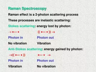
History of Raman scattering • 1923 • Predicted by Adolf Smekal • 1928 • Observe on particle • By means of sunlight • 1930 • Nobel Prize for Physics • 1960 • Laser • 1974 • SERS • Recently, nano-tech….. C. V. Raman, 1888-1970 /rɑːmən/
Vibration spectra of molecules Electronic transition (in VIS or UV) Energy Virtual state (Raman) Excited electric state Rotation transition (in microwave) Ground state Vibration transition (in infrared) Internuclear separation
Vibration spectra [Cu(pic)2].2H2O
Principle of Raman scattering The Vs is the Raman spectra.
Setup of Raman scattering system CCD camera Based on this simple scheme… • Embedded to microscopy • Embedded to SNOM • Embedded to optical tweezers • Embedded with time-resolved module • Embedded with FCS or TPM • Develop a portable device • …….. Energy monitor Laser UV-NIR spectrograph BS Notch filter or long pass filter polarizer Interference filter Specimen
What is SERS? • Surface-Enhanced Raman Scattering • The first time in 1974, • pyridine molecules • Electrochemically roughened silver surface • Electromagnetic contribution • the increase of the optical field intensity in the proximity of sharp points • Local surface plasmon resonance (LSPR) • Chemical effect • the mixing of the orbital of the adsorbed molecule and the metal atoms. • Example of electric field localization in colloids and sharp point samples. • The field intensity depends on the inter-particle distance and particle shape www.d3technologies.co.uk/en/10256.aspx
Principle of SERS So far, the theoretical understanding is not clear….. KatrinKneipp, HaraldKneipp, J. Phys.: Condens. Matter 14 (2002) N molecules with Metal particle (10~100nm) • Raman signal is too weak • Surface enhanced Raman scattering (SERS) technique can enhance Raman signal to 108 times • Electromagnetic effect and chemical effect between nanoparticles and molecules. N’ molecules with
How does SERS work? • (1) laser light incident on the metal substrate (2) plasmons excitation (3) light scattered by the molecule (4) Raman scattered light transferred back to plasmons and scattered in air (5) • The plasmon properties – such a wavelength and width of its resonance – depend on the nature of the metal surface and on its geometry. www.d3technologies.co.uk/en/10256.aspx
Lowest detection limitation • In 1977, Van Duyne and Jeanmaire and, independently, Albrecht and Creighton confirmed SERS experiment in 1974 • Surface effect + nanostructure effect • Prove the EM effect • SERS enhancement factors • Modest 103 to 105 • Dye 1010 to 1011 • 1995, effective cross section=(anti-Stocks)/(Stocks), to identify no resonance SERS cross section to 10-16 cm2 per molecule, and 1014 order of magnitude. ACCOUNTS OF CHEMICAL RESEARCH / VOL. 39, NO. 7, 2006
SERS-active substrates • Rough metal surface at nanometer • Gold/ silver colloid • Gold/ silver periodic nano-structures • Other nanostructures….. www.tedpella.com/gold_html/goldsols.htm
Applications of SERS • Light source: NIR/ VS/ UV • Subtracts: various metal nanostructures • Analytes: • Single molecule detection (R6G….) • Identification of a single DNA base molecule • Glucose sensor (J. AM. CHEM. SOC. 9 VOL. 125, NO. 2, 2003) • Protein • Cell
Assembly of Gold Nanostructured Films Templated by Colloidal Crystals and Use in SERS 4EC rough gold 5 after heating 500 C 6 glass wo metal 1gold NPs (25 nm) 2 no latex 3 only bulk BPE trans-1,2-bis(4-pyridyl)ethylene (BPE) J. Am. Chem. Soc. 2000, 122, 9554-9555
Single Molecule Detection Using SERS • Measured spectra of a single crystal violet molecule in aqueous colloidal silver solution using one second collection time and nonresonant near-infrared excitation show a clear “fingerprint” of its Raman features between 700 and 1700 cm-1. Spectra observed in a time sequence for an average of 0.6 dye molecule in the probed volume exhibited the expected Poisson distribution for actually measuring 0, 1, 2, or 3 molecules. Cite # 1138 Phys. Rev. Lett. 78, 1667 - 1670 (1997) Science ,1997, Vol. 275. no. 5303, pp. 1102 - 1106
Quantitative Simultaneous Multianalyte Detection of DNA by Dual-Wavelength SERS • Quantitative identification of specific DNA sequences in a mixture • Silver nanoparticles Angew. Chem. 2007, 119, 1861 –1863
Femtomolar Detection of Prostate-Specific Antigen: An Immunoassay Based on SERS and Immuno gold Labels Infrared reflection spectra of a DSNB-derived monolayer on gold before (spectrum A) and after (spectrum B) exposure to the anti-PSA tracer antibody. Anal. Chem. 2003, 75, 5936-5943
Demonstration of a SERS-based free PSA immunoassay. (A) SERS spectra, offset for clarity, acquired at various PSA concentrations. (B) Dose-response curve for free PSA in human serum. The dose-response curve was constructed by calculating the average reading of the response for 6-8 different locations on the surface of each sample, which typically varied by 10% (see text for further details). Anal. Chem. 2003, 75, 5936-5943
SERS in Local Optical Fields of Silver and Gold Nanoaggregatess From Single-Molecule Raman Spectroscopy to Ultrasensitive Probing in Live Cells Kneipp et al. Schematic of hyper-Raman and Raman scattering and surface-enhanced Raman and hyper-Raman spectra of crystal violet on silver nanoclusters, excitation 850 nm, 107 W/cm2. Stokes and anti-Stokes SERS spectra of crystal violet attached to isolated and aggregated gold nanospheres. Acc. Chem. Res. 2006, 39, 443-450
(a) Cells of a fibroblast cell line, NIH/3T3 (nonphagocytic) (left), and a macrophage cell line, J774 (phagocytic) (right), after uptake of gold nanoparticles; particle accumulations are visible as black dots inside the cells. Scale bars ) 20 ím. (b) Examples of SERS spectra acquired from NIH/3T3 cells after 3 h incubation with gold nanostructures, excitation wavelength 830 nm, 1 s collection time. Ex: proteins (1245 cm-1, 1267 cm-1 amide III, side chains Phe 1002 cm-1, Tyr 825 cm-1) and various nucleic acid constituents (e.g. 1580, 1575, 1098 cm-1) Acc. Chem. Res. 2006, 39, 443-450
In vivo tumor targeting and spectroscopic detection with surface-enhanced Raman nanoparticle tags NATURE BIOTECHNOLOGY VOLUME 26 NUMBER 1 JANUARY 2008
In vivo SERS spectra obtained from pegylated gold nanoparticles injected into subcutaneous and deep muscular sites in live animals. 5hr, N=4
Optical aggregation of metal nanoparticles in a microfluidic channel for surface-enhanced Raman scattering analysis 514.5 nm For tweezers, 830nm Lab Chip, 2009, 9, 193–195
Flow rate of 0.001ml/min 1s integration, 10s interval Dark field (DF) images and time series SERS spectra during an optical trapping process in the microfluidic channel. SERS measurements using a Y-shaped channel. Lab Chip, 2009, 9, 193–195
Advantages of SERS • Include all advantages of Raman spectroscopy • Fingerprint of molecules • Identify target in mixture • Living cell and the specimen contained water • Low concentration of biomolecue • Modification technique for those metal nanoparticles or other nanostructure with biomolecule • Integrated into bio-MEMS • Powerful probe for these applications with extreme low concentration.