Download
1 / 23
250 likes | 347 Views
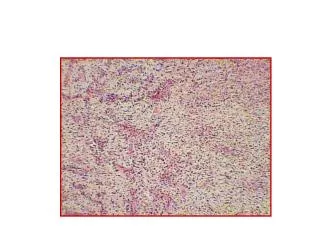
Periapical radiography. Main indications. Detection of periapical infection Assessment of the periodontal status After trauma to the teeth and associated alveolar bone Assessment of the presence and position of unerupted teeth. During endodontics

E N D
Main indications • Detection of periapical infection • Assessment of the periodontal status • After trauma to the teeth and associated alveolar bone • Assessment of the presence and position of unerupted teeth
During endodontics • Preoperative assessment and postoperative appraisal of apical surgery • Detailed evaluation of bone lesions • Implant evaluation
Ideal positioning requirements • The tooth under investigation and the film packet should be in contact or ,if not feasible, as close as possible • The tooth and the film packet should be parallel to one another
The film packet should be positioned with its long axis vertically for incisors and canines, and horizontally for premolars and molars with sufficient film beyond the apices to record the apical tissues The positioning should be reproducible
The X-ray tubehead should be positioned so that the beam meets the tooth and the film at right angles in both the vertical and the horizontal planes
Advantages of the paralleling technique • Geometrically accurate images are produced with little magnification, foreshortening or elongation • The shadow of the zygomatic buttress appears above the apices of the molar teeth
The horizontal and vertical angulations of the X-ray tubehead are automatically determined by the positioning device • There is no cone cutting • The radiographs are reproducible • The technique is useful for some patients with disabilities
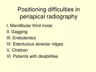
Disadvantages of the paralleling technique • Positioning of the film packet for the posterior teeth can cause gagging • The anatomy of the mouth sometimes makes the technique imposible, e.g a shallow, flat palate • The apices of the teeth can sometimes appear very near the edge of the film
The technique cannot be performed satisfactory using a short focal spot to skin distance because of the resultant magnification • The holders need to be autoclavable • Positioning the holders in the lower third molar region can be very difficult
Advantages of the bisected angle technique • Positioning of the film packet is relatively simple and comfortable for the patient • If the angulations are assessed correctly the image should be diagnosticable
Disadvantages • Incorrect vertical angulation results in foreshortening or elongation of the image • The shadow of the zygomatic buttress overlies the roots of the upper molars • Incorrect horizontal angulation results in overlapping of the crowns or cone cutting
The periodontal bone levels are poorly shown • It is not possible to obtain reproducible views • The buccal roots of the maxillary premolars and molars are foreshortened