Download
1 / 56
570 likes | 762 Views
Respiration + Vocal Fold Physiology. Feburary 6, 2014. Average Everydayness. Production exercise #1 is due today! I’m hoping to start grading it (and the DSP exercise) tonight. There will be a second production exercise, due before the break. Course project report #2 is due on Tuesday!

E N D
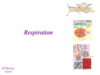
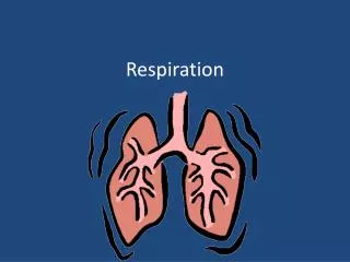
Respiration + Vocal Fold Physiology Feburary 6, 2014
Average Everydayness • Production exercise #1 is due today! • I’m hoping to start grading it (and the DSP exercise) tonight. • There will be a second production exercise, due before the break. • Course project report #2 is due on Tuesday! • Today: • The Wonderful World of the Larynx!
From the Bottom Up • All speech sounds require airflow. • The vast majority of sounds in the world’s languages use a pulmonic egressive airflow. • = out of the lungs • Questions to answer/consider: • How do we make air flow out of the lungs? • How does pulmonic airflow differ in breathing and in speech? • How does pulmonic airflow relate to language? • primarily: suprasegmentals (stress, F0)
The Machinery • The human torso (from the neck to the legs) has two major divisions: • The thorax • consisting of the heart and lungs • the “chest” • The abdomen • includes the digestive system and other interesting glands • the “belly”
The Thorax • The heart and the pulmonary system are enclosed by the thoracic cage. • the “rib cage” • Ribs are connected by cartilage to the sternum. • The intercostal muscles fill in the gaps between ribs... • and also cover the surfaces of the thoracic cage.
Connections • The thorax is split from the abdomen by a dome-shaped structure known as the diaphragm. • The lungs sit on top of the diaphragm. • Two membranes link the lungs to the ribs: • The visceral pleura covers the lungs. • The parietal pleura lines the inside of the thoracic cage.
Equilibrium • The linkage between the lungs and the rib cage makes: • The lungs are bigger than they would be on their own. • The rib cage is smaller than it would be on its own. • The linkage tends towards a natural equilibrium point. volume
Taking A Step Back • Air flows naturally from areas of high pressure to areas of low pressure. • Q: How do we make air pressure differences? • A: We take advantage of Boyle’s Law. • Boyle’s Law states that: • the pressure of the gas in a chamber is inversely proportional to the volume of gas in the chamber • The pressure of the gas can be increased or decreased by changing the volume of the chamber. • decreasing volume increases pressure • increasing volume decreases pressure
Inspiration • A normal breathing cycle begins with inspiration • “breathing in” • Air will flow into the lungs if... • the air pressure inside the lungs is lower than it is outside the lungs • Air pressure can be decreased inside the lungs by... • expanding the volume of the lungs. • Lung volume can be expanded: • In all three dimensions • With two primary muscle mechanisms
Expansion #1 • The vertical expansion of the thorax is primarily driven by the contraction of the muscles in the diaphragm. • This bows out the front wall of the abdomen. • Also: diaphragm contraction elevates the lower ribs. • expands the circumference of the thorax.
Expansion #2 • The thorax can also be expanded through the contraction of the external intercostal muscles. • Contraction of each intercostal muscle lifts up the rib beneath it. • Also pulls each rib forward with the sternum. • = expansion in the front-back dimension. sternum
Expansion #3 • The thorax can also be expanded through the contraction of the external intercostal muscles. • Contraction of the intercostals elevates the lower ribs more than the upper ribs • Lower ribs lift like a “bucket handle” • Expansion in the side-to-side dimension.
Expiration • Air flows out of the lungs whenever air pressure in the lungs is greater than external air pressure. • Note: technical term • alveolar pressure = air pressure inside the lungs • Alveolar pressure may be increased by decreasing lung volume. • Lung volume is decreased through both passive and active forces. • Normally, lungs contract after inspiration due to passive forces alone. • No muscular effort is necessary!
Passive Expiration • Thorax + lungs combo contracts back to its equilibrium point without any external impetus. • Relaxation pressure is inherent pressure on the lungs to revert back to the equilibrium point. • Note: relaxation pressure works both ways.
Active Expiration • Lung volume can be actively decreased by contracting a variety of muscles which: • Lower the ribs and/or sternum • thereby compressing the thorax in the front-to-back and side-to-side dimensions • Increase abdominal pressure • thereby driving the diaphragm upwards
Expiration #1 • The thoracic cage can be compressed by contracting the internal intercostals and the transversus thoracis. • These pull the ribs downward... • effectively the opposite action of contracting the external intercostals.
Expiration #2 • The most important muscles for active expiration increase pressure in the abdomen. • These include the rectus abdominis, the external and internal obliques, and the transversus abdominis. • Contracting these muscles drives in the abdomen... • and pulls down the sternum and lower ribs.
Expiration Dynamics • Technical term: the equilibrium point of thorax + lung volume is called the resting expiratory level. • At volumes above the resting expiratory level, the lungs will contract due to relaxation pressure alone. • ...although active expiration forces may contribute. • Below the resting expiratory level, the lungs will tend to expand due to relaxation pressure. • To continue expiration at this level, active expiration is necessary.
More Verbiage • Total lung capacity: volume of air in the lungs after a maximum inspiration. • Residual volume: amount of air that remains in the lungs after a maximum expiration. • Vital capacity: greatest amount of air that can be expelled from the lungs after a maximum inspiration. • = total lung capacity - residual volume • Functional residual capacity: volume of air contained in the lungs at the resting expiratory level. • Inspiratory capacity: maximum volume of air that can be inspired from the resting expiratory level. • = total lung capacity - functional residual capacity
Verbiage Diagram #1 residual volume total lung capacity vital capacity
Verbiage Diagram #2 functional residual capacity total lung capacity inspiratory capacity Note: FRC - RV only 35% of vital capacity
Keeping it Steady • The production of speech generally requires a continuous flow of air from the lungs. • A continuous flow of air requires constant alveolar pressure in the lungs. • Accomplishing this is tricky... • Because there is more relaxation pressure at the extremes (both high and low) of lung capacity. • Active inspiratory and expiratory forces have to dynamically compensate.
external air pressure expiratory pressure • Relaxation pressure changes from expiratory to inspiratory... • in going from maximum to minimum vital capacity inspiratory pressure constant pressure needed for utterance
Effort required to maintain constant alveolar pressure: effort initially requires inspiratory forces! active inspiratory pressure effort eventually requires expiratory forces active expiratory pressure
Electromyography (EMG) • The activity of inspiratory and expiratory muscles during continuous exhalation has been documented with electromyography (EMG) studies. • In EMG, an electrode is inserted into a particular muscle. • When that muscle contracts, it discharges an electrical signal (an action potential). • The voltage and timing of this discharge may be recorded through the electrode.
EMG Recordings Diaphragm External Intercostals Internal Intercostals Rectus Abdominis External Oblique Latissimus Dorsi inspiratory expiratory
Loudness • The intensity of an utterance is primarily determined by alveolar pressure. • Doubling alveolar pressure increases intensity by 9 -12 dB. • Louder utterances require a greater difference between alveolar and external air pressures • Louder utterances require more active expiratory force.
Differences • Conversational speech makes different demands on the respiratory system than either normal breathing or the production of a continuous vowel. • For instance, a normal breath cycle lasts about five seconds: • 40% of the cycle is devoted to inspiration • 60% of the cycle is devoted to expiration • In speech: • 10% of the cycle is devoted to inspiration • 90% of the cycle is devoted to expiration (i.e., talking)
Volume Differences • Normal breathing encompasses 35% - 45% of vital capacity. • Note: normally ends above the resting expiratory level. • Normal conversational speech encompasses 35% - 60% of vital capacity. • Loud speech usually starts at 60% - 80% of vital capacity. • and may end considerably above resting expiratory level. • Note: extremes of the vital capacity are not normally used, in either breathing or speech. • (requires too much muscular effort)
Modulating Airflow • During conversational speech, there are frequent demands for rapid changes in muscular pressure. • These changes are primarily required for differentiating between stressed and unstressed syllables. • Rapid modulations to airflow are primarily made by the internal intercostal muscles.
Lastly • Higher airflow from lungs = increase in F0 • The have-your-friend-punch-you-in-the-stomach experiment. • Increased F0 also contributes to stress. • However, F0 level is primarily determined by laryngeal activity... • which we’ll talk about next...
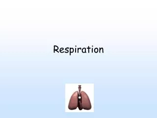
Where Were We? • Air squeezed out of the lungs travels up the bronchi... • Through the trachea (windpipe) • To a complicated structure called the larynx. • ...where phonation happens.
The Larynx • The larynx is a complex structure consisting of muscles, ligaments and three primary cartilages.
1. The Cricoid Cartilage • The cricoid cartilage sits on top of the trachea • from Greek krikos “ring” cricoid cartilage • It has “facets” which connect it to the thyroid and arytenoid cartilages.
2. The Thyroid Cartilage • The thyroid cartilage sits on top of the cricoid cartilage. • from the Greek thyreos “shield” • The thyroid cartilage has horns! • Both lower (inferior) and upper (superior) horns • The lower horns connect with the cricoid cartilage at the cricoid’s lower facet. • The upper horns connect to the hyoid bone.
Thyroid Graphic thyroid cartilage cricoid cartilage
Thyroid Angles • The two broad, flat front plates of the thyroid--the laminae--meet at the thyroid angle. • The actual angle of the thyroid angle is more obtuse in women. • ...so the “Adam’s Apple” juts out more in men.
3. The Arytenoid Cartilages • There are two arytenoid cartilages. • from Greek arytaina, “ladle” • They are small and pointy, and sit on top of the back side, or lamina, of the cricoid cartilage. arytenoid cartilages cricoid cartilage
The Vocal Folds • These three cartilages are connected by a variety of muscles and ligaments. • The most important of these are the vocal folds. • They live at the very top of the trachea, in between the cricoid and thyroid cartilages. • The vocal folds are a combination of: • The vocalis muscle • The vocal ligament • The vocal folds are enclosed in a membrane called the conus elasticus.
Vocal Fold View #1 • Just above the true vocal folds are the “false” (!) vocal folds, or ventricular folds. • The space between the vocal folds is the glottis.
Vocal Fold View #2 • The vocal ligaments attach in the front to the thyroid cartilage. • ...and in the back to the arytenoid cartilages. • The glottis consists of: • the ligamental glottis • the cartilaginous glottis
Things Start to Happen • Note that the arytenoid cartilages can be moved with respect to the cricoid cartilage in two ways. #1: rocking #2: sliding
The Upshot • The arytenoids can thus be brought together towards the midline of the body. • Or brought forwards, towards the front of the thyroid. • The rocking motion thus abducts or adducts the glottis. • The sliding motion shortens or lengthens the vocal folds. • Check out the arytenoids in action.
When the vocal folds are abducted: • air passes through the glottis unimpeded and voicelessness results. • The posterior cricoarytenoid muscles are primarily responsible for separating the arytenoid cartilages.
Voicing may occur when the vocal folds are adducted and air is flowing up through the trachea from the lungs. • Two muscles are primarily responsible for adducting the vocal folds. • The first is the lateral crico-arytenoid muscle.
Note that the lateral cricoarytenoid muscles only adduct the ligamental glottis. • The transverse arytenoid muscles pull together the arytenoid cartilages themselves. • Thereby closing the cartilaginous glottis.
The Consequences • The combined forces drawing the vocal folds towards each other produce adductive tension in the glottis. • Adductive tension is increased by: • lateral cricoarytenoid muscles • transverse arytenoid muscles • Adductive tension is decreased by: • posterior cricoarytenoid muscles • Adduction vs. abduction determines whether or not voicing will occur. • But we can do more than just adduce or abduce the vocal folds...
Controlling F0 • Question: why do women have a higher F0 than men? • A: Shorter vocal folds open and close more quickly. • In men: • Ligamental glottis 15.5 mm • Cartilaginous glottis 7.5 mm • Total glottis length 23 mm • In women: • Ligamental glottis 11.5 mm • Cartilaginous glottis 5.5 mm • Total glottis length 17 mm
Factor Two • F0 also depends on the longitudinal tension in the vocal folds. • I.e., tension along their length, between the thyroid and arytenoid cartilages. • Higher tension = higher F0 • Lower tension = lower F0 • Q: How can we change longitudinal tension in the larynx?
A: We can rotate the thyroid cartilage up and down on its connection with the cricoid cartilage. • ...like the visor of a knight’s helmet. • This either stretches or relaxes the vocal folds.