Download
1 / 10
100 likes | 220 Views
Pages 52 to 54. Microscopes. Types of microscope. Compound Light Visible light is used to illuminate the specimen Glass lens bend the light to magnify the image Magnifies up to 1,000X Limited resolution Able to view living things

E N D
Pages 52 to 54 Microscopes
Types of microscope • Compound Light • Visible light is used to illuminate the specimen • Glass lens bend the light to magnify the image • Magnifies up to 1,000X • Limited resolution • Able to view living things • Various techniques can be used to differentiate structures – dyes, fluorescence
Types of Microscopes • Electron • Beam of electrons to view image • Much greater magnification • Better resolution • Cannot view living things • Transmission Electron Microscope (TEM) • Shows internal detail • Scanning Electron Microscope (SEM) • Gives 3-dimensional image of surface Scanning ↑ Transmission ↓
10 m Human height 1 m Length of some nerve and muscle cells 0.1 m Unaided eye Chicken egg 1 cm Frog egg 1 mm 100 µm Most plant and animal cells Light microscope 10 µm Nucleus Most bacteria Mitochondrion 1 µm Electron microscope Smallest bacteria 100 nm Viruses Ribosomes 10 nm Proteins Lipids 1 nm Small molecules Atoms 0.1 nm
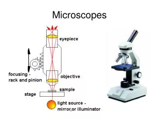
Parts of the Microscope • Eye piece or ocular lens • What you look thru • Magnifies 10X • Revolving nosepiece • Allow you to change the objective lens • Objective lenses • Magnify the image • Scanning Power 4X • Low Power 10X • High Power 40X • Stage • Holds the slide • Mechanical stage makes adjustments easy
Parts of the Microscope • Light Source • Illuminates the object • Diaphragm • Adjustable lever below stage • Regulates the amount of light passing through the image (too bright – close / too dark – open) • Base • Supports the microscope • What you hold to carry it • Coarse adjustment • Focus in large increments • Only use with scanning & low • Fine adjustment • Focus in small increments
Start on the scanning objective Use the coarse adjustment to bring object into view Use fine adjustment to make clear Move object to center of field of view Switch objectives use fine adjustment to make clear NEVER use coarse adjustment on high power How to focus a microscope
How to make a wet mount slide • Place a drop of water onto slide • Place object in water • Use a probe to slowly lower the cover slip onto the slide • Try to avoid air bubbles
Total magnification = magnification of the ocular lens X the magnification of the objective lens • Scanning Power Total Magnification: • 10 X 4 = 40 • Low Power Total Magnification: • 10 X 10 = 100 • High Power Total Magnification: • 10 X 40 = 400 How to calculate total magnification
Draw to the best of your ability • No scribbling • No lines/dot • Label the image • What are you looking at? • Document the total magnification that you are using at the time. • 40X, 100X or 400X When you Draw an object