Download

1 / 50
530 likes | 1.38k Views
Airway Management Part III. RET 2275 Respiratory Care Theory 2. Tracheostomy Tubes. A long-term airway placed through an incision made between the 2 nd and 3 rd tracheal rings and inserted directly into the trachea. Tracheostomy Tubes. Indications for tracheostomy:

E N D
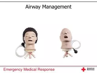
Airway ManagementPart III RET 2275 Respiratory Care Theory 2
Tracheostomy Tubes • A long-term airway placed through an incision made between the 2nd and 3rd tracheal rings and inserted directly into the trachea
Tracheostomy Tubes • Indications for tracheostomy: • Airway obstruction due to the following: • Inflammatory disease • Benign laryngeal pathology, e.g., webs, cysts, papilloma) • Malignant laryngeal tumors • Laryngeal trauma or stenosis • Tracheal stenosis • Pulmonary toilet • Obstructive sleep apnea
Tracheostomy Tubes • Advantages over prolonged translaryngeal intubation: • Eases airway care and suctioning • Eliminates the ongoing risks of oral, nasal, pharyngeal, and most laryngeal complications of translaryngeal intubation • Reduces risk of tracheal extubation • Eases tube reinsertion • Facilitates oral communication and speech • Improves oral, nasal, and facial hygiene
Tracheostomy Tubes • Advantages over prolonged translaryngeal intubation: • Raises patient comfort level • Improves patient appearance • Facilitates nursing care of the overall airway • Improves patient mobility • Eases disposition to long-term care facility • Less airway resistance
Tracheostomy Tubes • Most experts agree that patients requiring ET intubation for more than 7 days should have a tracheostomy • Some evidence indicates that a tracheostomy performed early, i.e., within 3 days of intubation, may decrease the risk for pneumonia, the length of mechanical ventilation, and the length of stay in the ICU
Tracheostomy Tube • Outer Cannula; primary structural unit of the tube, to which is attached the cuff and flange • Flange; prevents tube slippage into the trachea and provides means to secure the tube to the neck • Inner Cannula; cannula within the outer cannula that can be removed for routine cleaning – can be locked in place
Tracheostomy Tube • Cuff; seals off the lower airway, either for protection from aspiration or to provide positive pressure ventilation – inflation tube (aka: pilot tube) leads from the cuff to a pilot balloon and spring loaded valve. • Tie strings; stabilizes the tube at the stoma site - attached to the flange and is tied around the neck
Tracheostomy Tube • Obturator; placed within the outer cannula with its tip extending just beyond the far end of the tube – minimizes mucosal trauma during insertion • Radiopaque Indicator; helps confirm tube position on radiograph
Airway Trauma with Tracheal Tubes Laryngeal Lesions Glottic edema Vocal cord inflammation Both are transient changes that occur as a result of pressure from the ETT, or trauma during intubation Symptoms include hoarseness and stridor Primarily a concern after extubation and can worsen over 24 hours
Airway Trauma with Tracheal Tubes Laryngeal Lesions Laryngeal and vocal cord ulcerations May cause hoarseness after extubation Symptoms usually resolve spontaneously Vocal cord polyps and granulomas Develop more slowly – taking weeks or months Symptoms include: Difficulty swallowing Hoarseness Stridor May have to be removed surgically
Airway Trauma with Tracheal Tubes Laryngeal Lesions Vocal cord paralysis Symptoms may resolve within 24 hours If obstructive symptoms continue, tracheotomy may be indicated Laryngeal stenosis Normal tissue is replaced by scar tissue, which causes stricture Symptoms include stridor and hoarseness Surgical correction is usually required
Airway Trauma with Tracheal Tubes Tracheal Lesions Granulomas Circumscribed mass of cells (mainly histiocytes) normally associated with the presence of chronic infecton or inflammation Tracheomalacia Softening of the cartilaginous rings, which causes collapse of the trachea during inspiration
Airway Trauma with Tracheal Tubes Tracheal Lesions Tracheal stenosis Narrowing of the lumen of the trachea, which can occur a fibrous scarring causes the airway to narrow With ETT, most often occurs at the site of the cuff With tracheostomy tubes, occurs at the cuff, tube tip, or stoma site (most common)
Airway Trauma with Tracheal Tubes Tracheoesophageal fistula A direct communication between the trachea and esophagus Development is related to sepsis, malnutrition, tracheal erosion from the cuff and tube and esophageal erosion from nasogastric tubes
Airway Trauma with Tracheal Tubes Tracheal Lesions Tracheoinnominate fistula Occurs when a tracheostomy tube causes tissue erosion through the innominate artery Results in hemorrhage and, in most cases, death
Prevention of Tracheal Lesions Limit Tracheal Tube Movement Sedation Nasotracheal tube intubation Swivel adaptors for equipment attached to tracheostomies Tracheostomy collars instead of T-tubes
Prevention of Tracheal Lesions Selection of correct size airway Limit cuff pressure Maintain sterile technique when caring for tracheal tubes to limit infection Good and regular tracheostomy care
Providing for Patient Communication • To help facilitate communication between healthcare givers and patients who cannot speak because of having an endotracheal or standard tracheostomy tube in place, various devices can be utilized
Providing for Patient Communication Communication Board
Talking Tracheostomy Tube Provides a separate inlet for compressed gas, which escapes above the tube allowing phonations
Passy-Muir Speaking Valves A one-way valve on the external end of the tracheostomy tube that allows the patient to inhale through the tube and exhale through the larynx The cuff on the tracheostomy tube must be deflated)
Passy-Muir Valve • Candidates for PMV • Awake and alert tracheostomized (ventilator or non-ventilator dependent patients) – adult, pediatric and neonatal
Passy-Muir Valve • Benefits • Tracheostomized and ventilator dependent patients can produce clearer speech • Improved swallowing due to increased pharyngeal/laryngeal sensation decreasing the need for tube feeding • Decreased need for suctioning by enabling the patient to produce a stronger, effective cough
Passy-Muir Valve • Benefits • Decreased aspiration due to increased pharyngeal/laryngeal sensation • Improved weaning by improving physiologic PEEP, which can improve oxygenation • Reduces decannulation time by allowing the patient to begin to adjust to a more normal breathing pattern through the upper airway • Decreased length of stay
Passy-Muir Valve • Contraindications • Unconscious and/or comatose patients • Inflated tracheostomy tube cuff • Foam filled cuffed tracheostomy tube • Severe airway obstruction which may prevent sufficient exhalation • Thick and copious secretions • Severely reduced lung elasticity that may cause air trapping • This device is not intended for use with endotracheal tubes
Ensuring Adequate Humidity Artificial airways (ETT/Tracheostomy tubes) bypass the normal humidification, filtration, and heating function of the upper airway, which can cause: Secretions to thicken Impairment of ciliary function Impairment of mucocilary clearance Secretion retention
Ensuring Adequate Humidity Heated humidification Large volume nebulizer Heat and moisture exchanger (HME)
Minimizing Nosocomial Infection Patient with tracheal airways (ETT/Tracheostomy tube) are susceptible to bacterial colonization and infection of the lower respiratory tract Minimize by: Consistently wash hands before and after each patient contact Adhering to sterile technique during suctioning Use only aseptically clear or sterile respiratory equipment for each patient
Care of the Tracheostomy Tube Tracheostomy tubes require daily care Assemble and check equipment PPE; masks, goggles, gown, gloves Suction equipment Oxygen, manual resuscitator Tracheostomy cleaning kit
Care of the Tracheostomy Tube Tracheostomy tubes require daily care Suction the patient Clean the inner cannula Remove the inner cannula and place in the basin with hydrogen peroxide to soak Insert a disposable inner cannula if on mechanical ventilator Clean inside and outside of cannula with a brush and rinse with sterile water Allow to dry
Care of the Tracheostomy Tube Tracheostomy tubes require daily care Clean the stoma site Remove the patient’s gauze dressing and discard in a biohazard container Using cotton-tipped applicators, or sterile gaze dipped in a hydrogen-sterile water solution to clean under the flange and around the stoma Using a sterile gauze dipped only in sterile water, rinse stoma site
Care of the Tracheostomy Tube Tracheostomy tubes require daily care Clean the stoma site (cont.) Place a clean gaze under the flange Do not cut gauze for this purpose as fibers may loosen and become caught in the stoma Use precut gauze or folded 4 x 4 gauze pads Change ties Replace clean inner cannula
Extubation / Decannulation For most patients, tracheal intubation is a temporary measure The process of removing an artificial tracheal airway is called extubation
Assessing Readiness to Extubate Original need to for the artificial airway no longer exists Able to protect airway Presence of a gag reflex Able to manage secretions Cough strength Quantify and thickness of secretions Patency of the upper airway
Orotracheal / Nasotracheal Tubes Procedure Assemble Equipment Suction apparatus O2 / Lg. volume nebulizer Resuscitator/mask SVN with racemic epinephrine and NSS Intubation tray
Orotracheal / Nasotracheal Tubes Procedure Suction ETT and pharynx to above the cuff After use, prepare rigid tonsillar (yankauer) suction tip Oxygenate the patient well after suctioning Give 100% oxygen for 1 – 2 minutes Deflate the cuff
Orotracheal / Nasotracheal Tubes Procedure Remove the tube Method 1 Give a large breath with manual resuscitator and remove tube at peak inspiration Method 2 Ask the patient to take a deep breath and cough, pull the tube during the expulsive expiratory phase
Orotracheal / Nasotracheal Tubes Procedure Apply appropriate oxygen and humidity Patients who have been receiving mechanical ventilation may still require oxygen therapy, usually a higher FIO2 If humidity therapy is indicated, most clinicians suggest a cool mist aerosol, which helps reduce the swelling that normally occurs after extubation Encourage the patient to cough
Orotracheal / Nasotracheal Tubes Procedure Assess/Reassess the patient Air movement Auscultate and listen for good air movement; stridor or decreased air movement after extubation indicates upper airway problems RR, HR, BP, SpO2 ABG as needed
Orotracheal / Nasotracheal Tubes Complications The most common problems after extubation are hoarseness, sore throat, and cough – these are benign and will resolve with time Laryngospasm, a rare but serious complication associated with extubation, is usually transient and treatable with high FiO2 and application of positive pressure. If it persists, a neuromuscular blocking agent may have to be given, which will necessitate manual ventilation or reintubation
Tracheostomy Tube Removal There are several approaches to removing tracheostomy tubes (“decannulation”) – the method used will be depend on the patient’s needs Abrupt - removed in one step Weaning Fenestrated tubes Progressively smaller tubes Tracheostomy buttons
Fenestrated Tracheostomy Tube The fenestrated tracheostomy tube has a removable inner cannula. However, the outer cannula has a hole in its posterior wall - a fenestration With the inner cannula removed and the cuff deflated, the patient may breathe through the upper airway, via the fenestration - allowing for increasing use of the upper airway
Fenestrated Tracheostomy Tube In the event mechanical ventilation is required, the cuff can be inflated and inner cannula replaced; in this configuration, the tube performs like a regular cuffed tracheostomy tube
Progressively Smaller Tubes A second airway weaning technique is to use progressively smaller and smaller tracheostomy tubes Indicated in patients who airway is too small for a fenestrated tube May facilitate better healing of the stoma Problems Increases in airway resistance May impair coughing Smaller tubes may result in the curve of the tube impacting the posterior tracheal wall
Tracheostomy Button Used to maintain a tracheal stoma
Tracheostomy Button Fits from the skin to just inside the anterior wall of the trachea Avoids added resistance to the airway Use: Relieving airway obstruction Removing secretions
Tracheostomy Button Has an adaptor for provision of IPPB or mechanical ventilation An optional one-way valve on the external end of the button allows for speech
Assessment After Decannulation Vocal cord evaluation Abnormalities can result in aspiration or acute airway obstruction Symptoms Stridor Retractions Inability to feel airflow through the upper airway Have a replacement tracheostomy tube and suctioning equipment available