Download
1 / 73
750 likes | 769 Views
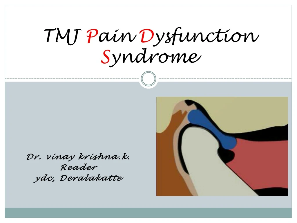
TMJ P ain D ysfunction S yndrome. Dr. vinay krishna.k . Reader ydc , Deralakatte. Temporomandibular disorder(s) (TMD) or temporomandibular joint (TMJ) syndrome is the most common cause of facial pain after toothache. SYNONIMS. TMJ disease or TMJ syndrome.

E N D
TMJ Pain Dysfunction Syndrome Dr. vinaykrishna.k. Reader ydc, Deralakatte
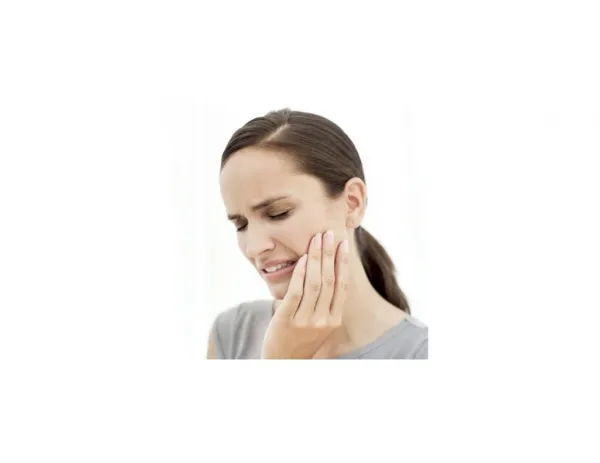
Temporomandibular disorder(s) (TMD) or temporomandibular joint (TMJ) syndrome is the most common cause of facial pain after toothache.
SYNONIMS TMJ disease or TMJ syndrome. Costen syndrome after Dr. James Costen (1934). Temporomandibular disorder (TMD) by American Academy of Orofacial Pain (AAOP). Temporomandibular joint disorder (TMJD) by National Institute of Dental and Craniofacial Research (NIDCR). Myofacial Pain Dysfunction Syndrome (MPDS)
AAOP classification TMD broadly divided into 2 syndromes: (1) Muscle-related TMD (myogenous TMD), sometimes this is called TMD secondary to myofacial pain and dysfunction (MPD), and (2) Joint-related (arthrogenous) TMD, that is TMD secondary to true articular disease.
Myogenous TMD is more common. Multifactorial,in its pure form, it lacks apparent destructive changes of the TMJ on radiograph and can be caused by multiple etiologies such as bruxism and daytime jaw clenching in a stressed and anxious person.
Arthrogenous TMD can be further specified as disk displacement disorder, chronic recurrent dislocations, degenerative joint disorders (DJDs), internal derangements, systemic arthritic conditions, ankylosis, infections, and neoplasia.
predilection Sex: TMD primarily affects women, more so young women. The male-to-female ratio is 1:4. Age: Highest incidence is among young adults, especially women aged 20-40 years.
Signs &Symptoms • PAIN. • Click, pop, and snap,Facialpain • Limited jaw opening(Trismus) and locking episodes. • Headaches. • Other- otalgia, neck pain and/or stiffness, shoulder pain, and dizziness. About one third of these patients have a history of psychiatric problems. • 60% spontaneous.
Internal Derangements • Tenderness to palpation • Pain with movement • Audible click
Degenerative Joint Disease Pain with joint movement Crepitus over joint Flattened condyle Osteophyte formation
Diagnosis • History • Clinical evaluation • Investigations • Radiographs • Ultrasound • CT • MRI • Diagnostic arthroscopy
MANAGEMENT • Medical Care: • Most TMDs are self-limiting and do not get worse. • Ibuprofen and naproxen are commonly used NSAIDs • muscle relaxants are diazepam, methocarbamol, and cyclobenzaprine • Tricyclic antidepressants- Amitriptyline and nortriptyline
Botulinum toxin is used both as a single treatment (Schwartz and Freund, 2002) and in conjunction with arthrocentesis (Freund and Schwartz, 2003).
Occlusal splints • 2 groups— • Anterior repositioning splints and • Autorepositional splints (muscle splints)
Surgical Care • Arthrocentesis • Arthroscopic surgery • Open surgery • disk repositioning and plication • disk removal • Myrhaug technique(1951)- resecting the temporal condyle, creates a permanent and reducible chronic dislocation of the joint. • Arthroplasty
OTHER METHODS • Physical therapy. • electromyographic (EMG) biofeedback • Friction massage • Ultrasonic treatment • Transcutaneous electronic nerve stimulation(TENS) • Cognitive-behavioral treatment. • Psychological treatment.
Subluxation • Condylarsubluxationis an incomplete joint the articular surfaces maintain partial contact and the condyle is able to return to the glenoidfossa voluntarily or aided by self manipulation. • The condition is related to internal derangement as the open, incomplete and transient dislocation
Dislocation • Dislocation is a complete separation of the articular surfaces with fixation in an abnormal position. • Anterior dislocation of the condyle in which the normal anatomic relationships within the joint have been completely disrupted occurs with the condyle displaced and fixed anterior to the articular eminence.
The key terms used include hypermobility, acute dislocation, long dislocation, recurrent dislocation and habitual dislocation. • Subluxation is substituted for the term dislocation where dislocation is incomplete . • luxation and dislocation are synonymous.
Dislocation can occur as a single acute event or as chronic recurring episodes. • Dislocations which take place repeatedly and which last for short or long interval are referred to as Recurrent dislocation. • A dislocation that remains locked anteriorly for several days years is an old or long-standing dislocation. • The term choric dislocation is most appropriately used in those cases where the patient is able dislocate and reduce at will.
Pathophysiology • Acute anterior dislocation is precipitated by either intrinsic or extrinsic trauma. • A wide yawn is a frequent cause of spontaneous (intrinsic) dislocation. • Other forms of intrinsic events such as vomiting,singinglaughing,screaming, wide biting,&seizures.
Extrinsic traumatic dislocation is due to violence which forces the condyle out of the fossa. • External force such as a blow to themandible,usually with the mouth in an open position, can result in mandibular dislocation. • Manipulation of the jaw during intubation for general anaesthesia, endoscopy & dental extraction is another extrinsic cause.
Laxity of ligaments& capsule &abnormalities of skeletal form are predisposing factors in both acute & chronic forms of dislocation. • Looseness of the capsule and ligaments can occur from inadequate healing after injuries, hypermobility& from longstanding degenerative joint disease.
Occlusal abnormalities and loss of vertical dimension from loss of teeth can also contribute to laxity & to the occurrence of recurrent dislocation. • Acute dislocation found the commonest cause to be a blow on the chin with the mouth open in males and dental extractions in females.
The activity and condition of the ligaments associated with the temporomandibular joint are important considerations in dislocation. • The tempromandibular ligament & capusle remained taut in all mandibular movements and mantained the mandible in articulation the cranial base. • Opening movements of the mandible caused the stylomandibular ligaments and sphenomandibularto become slack and folded.
Hypermobility • Hypermobility ofthis joint is characterized by excessive anterior movement of the condyle at maximum mouth opening without strain or symptoms. • Hypermobility, subluxation, and dislocation of the temporomandibular joint are interelated conditions, and hypermobility is likely a predisposing factor.
Systemic Hypermobility • Familial hypermobility syndromes loose-jointed individuals with articular symptoms comprise a very heterogeneous group. • In the EhlersDanlos syndrome the degree of hypermobility and the incidence of dislocation are closely related. • In this condition dislocations of the temporomandibular joint are often recurrent • EhlersDanlos syndrome the incidence of temporomandibular joint dislocations was 3.3%
Occlusal factors • Long-term overclosure and loss of physiologic vertical dimension secondary to loss of dentition can Contribute to subluxation & dislocation. • The mechanism of this is thought to be that overclosure produces stretching and loosening of joint ligaments and joint laxity can then lead to subluxation.
Asymmetry of the condylar position due to mandibularmalposition may be caused by occlusal interferences. • Occlusal disturbances may also be related to Bruxism. • Recurrent dislocation occurs in which extractions of bilateral distoangular, palatally inclined maxillary third molars eliminated the mandibular dislocation.
Drug-associated dislocation • Spontaneous dislocation of the mandible due to extrapyramidal reactions to prochlorperazine. • Left facial weakness and wild facial contortions occurred, followed by a unilateral mandibular dislocation.
Psychogenic dislocation • Hysteria can be the cause of habitual dislocation of the mandible. • Ligaments are lax and repetitious subluxation or dislocation can easily occur. • It is important to recognize early that habitual dislocation may be the presenting feature of an underlying psychiatric disturbance. • The degree and duration of the disability are out of all Proportion to the severity of the injury.
Diagnosis • A thorough History & physical examination is important to evaluate properly all categories of dislocation. • It is important to determine the cause & onset of the dislocation. • A spontaneous intrinsic dislocation only occurs in an anterior direction. • Acute, initial spontaneous and extrinsic traumatic anterior dislocations are treated differently from chronic repetitive dislocation.
A prior history of local joint laxity, internal derangements,&other temporomandibular joint disorders will influence the outcome of treatment and must be ascertained in evaluating the past history. • Neurologic & mucoskeletal disorders such as Parkinson's disease & epilepsy and other systemic disorder of hypermobility are important to recognize.
Clinical examination • Spontaneous dislocation from a wide yawn is often bilateral,but a blow to the chin with the mouth open usually create a unilateral dislocation. • Bilateral dislocation is associated with pain, inability to close the mouth,tensemasticatory muscles, difficulty with speech, excessive salivation a protruding chin and open bite. • The lateral pole of the condyle produces a characteristic protuberance anterior to and below the articular eminence which can usually be seen and palpated.
Unilateral dislocation is characterized by the mandible swung away from the side of dislocation. • The Devation produces a lateral cross and Open bite on the contra lateral side. • Palpation of the muscles and joints is a valuable aid to diagnosis. • Tenderness in the joint may indicate a fracture,where as tenderness in the temporal fossa is more characteristic of dislocation.
Radiographic examination • Plains flims such as transcranial radiograph & lateral tomograms are important in the idenfication & documentation of dislocation. • Arthrographic studies with recurrent dislocation have enlabed a differenation to be made between meniscotemporal & meniscocondylar types • MRI and CT scanning would to useful to identifying ligament and capsular tears and stretching. • Eletromyographic studies in dislocation and subluxction provide valuable information. DILOCATION WITH CONDYLE ANTERIOR TO DISK & EMINENCE
Non surgical Treatment • The initial acute the longstanding & the chronic recurring dislocations of the mandible require different treatments. • The acute dislocation needs immediate attention for relief of pain and anxiety and to minimize damage to the joint structure. • Reduction and immobilization for 4 weeks will allow damage ligaments, capsule, and disk to heal. • However in chronic case, Immobilization does nothing correct the problem of an unstable joint
The major problem to overcome in all dislocation is muscle contraction. • Treatment is different for troublesome repetitive dislocation where the etiology is psychologic compared with systemic hyermobility without psychologic implications.
Acute dislocation • Initial treatment is aimed at reducing tension, anxiety, and muscle spasm by using the simplest methods. • A tranquillizer or sedative may aid in gaining then relaxtion needed also pressure and message over coronoid processes can also benefit. • An impressive simple technique is used by injecting local anesthetic is injected into the depression in the glenoidfossa left by the dislocated condyle.
Manipulation is the next step. • Hippocrates remains an effective way to manipulate and reduce the dislocated mandible. • A common method currently used has the operator standing in front of the patient who is sitting with the head supported. • Thumbs are wrapped in gauze and placed on the occlusal surfaces of the mandibular molars or alveolar ridges. • The lower border of the mandible is grasped with the fingers and the patient is encouraged to relax and open in the direction of the dislocation. • By pressing firmly on the molars and elevating anteriorly with simultaneous backward pressure the condyle is relocated.
Yurino's Method • Places the patient in a supine position without a pillow. • The patient is encouraged to relax completely while the operator stands near the patient's head and holds the body of the mandible from the opposite side. • The patient is asked to open and close the mouth and, although it is difficult to do so,it is important for the patient to attempt this alone.
The operator moves the mandible up and down in phase with the patient's opening and closing movements. • The operator thenlocates the dislocated condyle with his thumb & simultaneously with the patient's closing motion pushes it completely downward while moving the body of the mandible upward. • By this procedure the condyle moves over the articular eminence and slips into the fossa.
Longstanding dislocation • The difficulty in reducing mandibular dislocation increases proportionately with time Muscle relaxation and manipulation are usually successful if carried out immediately or within a few hours. • Reduction by forcing the mandible downward with the thumbs in the molar region and simultaneous upward tiliting of the chin was tried first. • Condylectomy was the preferred method.
Nonsurgical treatment of recurrent dislocation Physical therapy: • The use of isometric exercises to improve opening and closing patterns is most important. • Synchronized isometric contraction exercises of masticatory opening muscles and their antagonists should be performed on a regular basis.
Isometric exercise similar to that described by Poswillois very helpful. • This relatively simple exercise trains the suprahyoid muscles to stabilize the mandible and reduce forward movement of the condyle in the early opening phase. • The exercise should be carried out several times a day for 4 weeks until dislocations are no longer a problem. • Then the exercise should be done indefinitely once or twice a day to maintain the stability and to prevent a return to paranormal function.
Symptomatic treatment • Patients with subluxation and dislocation often suffer arthralgia & myalgia and symptomatic treatment is necessary. • Analgesics and nonsteroidal anti-inflammatory drugs will relieve locomotor system pain whether in the joint, bone, tendon, ligament, or muscle. • Muscle relaxants and tranquillizers are useful. • An injection of a steroid such as methylprednisone gives excellent results in persistent synovitis in the hypermobility syndrome. • Long-acting corticosteroids should be avoided as they may lead to connective tissues atrophy and weakening of collagenous tissue, which may contribute to increasing joint laxity.
Occlusal treatment • Occlusal disturbances, such as cuspalinterfernces and non occlusion due missing teeth with loss of vertical support, should be corrected to prevent their contributing to the instability of the joint. • However,appliances can be useful in those individuals with coexisting internal derangement of the disk, bruxism, and muscle hyperactivity.