Download
1 / 79

E N D
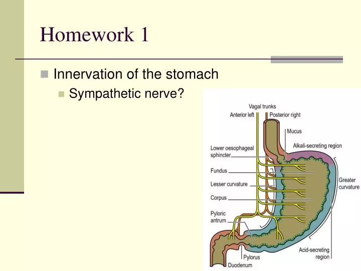
Homework 1 • Innervation of the stomach • Sympathetic nerve?
A, Parasympathetic. Dashed lines indicate the cholinergic innervation of striated muscle in the esophagus and external anal sphincter. Solid lines indicate the afferent and preganglionic efferent innervation of the rest of the gastrointestinal tract. • B, Sympathetic. Solid lines denote the afferent and preganglionic efferent connections between the spinal cord and the prevertebral ganglia. Dashed lines indicate the afferent and postganglionic efferent innervation. • CG, celiac ganglion; IMG, inferior mesenteric ganglion; SMG, superior mesenteric ganglion. Gastrointestinal Physiology, Seventh Edition. LEONARD R. JOHNSON. 2007, Mosby, Inc.
Homework 2 • Ionic mechanism of spike potential (Action potential): • Depolarization: Ca2+ influx? L-type Ca2+ current L-type Ca2+ channels provide the Ca2+ influx that initiates contraction. Blockade of Ca2+ channels reduces the duration and amplitude of electrical slow waves in many muscles and blocks generation of action potentials. Horowitz B, Ward SM, Sanders KM. Annu Rev Physiol. 1999;61:19-43.
Gastrointestinal Physiology (Part 2) Xia Qiang, PhD Department of Physiology Zhejiang University School of Medicine Email: xiaqiang@zju.edu.cn
Pancreatic juice • pH 7.8~8.4 • ~1500 ml/day • Isosmotic • Components: • Pancreatic digestive enzymes: secreted by pancreatic acini • Sodium bicarbonate: secreted by small ductules and larger ducts
At low magnification At higher magnification
Secretion of bicarbonate ions • Secreted by the epithelial cells of the ductules and ducts that lead from acini • Up to 145mmol/L in pancreatic juice (5 times that in the plasma) • Neutralizing acid entering the duodenum from the stomach
Secretion of pancreatic digestive enzymes Carbohydrates -- Pancreatic amylase Pancreatic lipase Fat Cholesterol esterase Phospholipase Trypsinogen Proteins Chymotrypsinogen Procarboxypolypeptidase Proelastase
Starches Pancreatic amylase Maltose and 3 to 9 glucose polymers
Trypsin Inhibitor • Inhibits the activity of trypsin and thus guards against the possible activation of trypsin and the subsequent autodigestion of the pancreas
Regulation of pancreatic secretion • Basic stimuli that cause pancreatic secretion • Ach • Cholecystokinin: • Secreted by I cells • Stimulates the acinar cells to secrete large amounts of enzymes • Secretin: • Released by S cells • Acts primarily on the duct cells to stimulate the secretion of a large volume of solution with a high HCO3- concentration
Stimulation of protein secretion from the pancreatic acinar cell. A, The pancreatic acinar cell has at least two pathways for stimulating the insertion of zymogen granules and thus releasing digestive enzymes. ACh and CCK both activate Gα , which stimulates PLC, which ultimately leads to the activation of PKC and the release of Ca . Elevated [Ca ] also activates calmodulin (CaM), which can activate protein kinases (PK) and phosphatases (PP). Finally, VIP and secretin both activate Gα , which stimulates adenylyl cyclase (AC), leading to the production of cAMP and the activation of PKA. B, Applying a physiological dose of CCK (i.e., 10 pM) triggers a series of [Ca ] oscillations, as measured by a fluorescent dye. However, applying a supraphysiological concentration of CCK (1 nM) elicits a single large [Ca ] spike and halts the oscillations. Recall that high levels of CCK also are less effective in causing amylase secretion.
In addition to protein, acinar cells in the pancreas secrete an isotonic, plasma-like fluid. Stimulation of isotonic NaCl secretion by the pancreatic acinar cell. Both ACh and CCK stimulate NaCl secretion, probably through phosphorylation of basolateral and apical ion channels. The rise in [Cl ] produced by basolateral Cl uptake drives the secretion of Cl down its electrochemical gradient through channels in the apical membrane. As the transepithelial voltage becomes more lumen negative, Na moves through the cation-selective paracellular pathway (i.e., tight junctions) to join the Cl secreted into the lumen. Water also moves through this paracellular pathway, as well as through aquaporin water channels on the apical and basolateral membranes. Therefore, the net effect of these acinar cell transport processes is the production of an isotonic, NaCl-rich fluid that accounts for ∼25% of total pancreatic fluid secretion.
Regulation of pancreatic secretion • Phases of pancreatic secretion: A meal triggers cephalic, gastric, and intestinal phases of pancreatic secretion • Cephalic Phase • Gastric Phase • Intestinal Phase
Three phases of pancreatic secretion. A, During the cephalic phase, the sight, taste, or smell of food stimulates pancreatic acinar cells, through the vagus nerve and muscarinic cholinergic receptors, to release digestive enzymes and, to a lesser extent, stimulates duct cells to secrete HCO and fluid. The release of gastrin from G cells is not important during this phase. During the gastric phase, the presence of food in the stomach stimulates pancreatic secretions'primarily from the acinar cells'through two routes. First, distention of the stomach activates a vagovagal reflex. Second, protein digestion products (peptones) stimulate G cells in the antrum of the stomach to release gastrin, which is a poor agonist of the CCK receptors on acinar cells. B, The arrival of gastric acid in the duodenum stimulates S cells to release secretin, which stimulates duct cells to secrete HCO and fluid. Protein and lipid breakdown products have two effects. First, they stimulate I cells to release CCK, which causes acinar cells to release digestive enzymes. Second, they stimulate afferent pathways that initiate a vagovagal reflex that primarily stimulates the acinar cells through M cholinergic receptors.
Acute pancreatitis • Acute pancreatitis is sudden swelling and inflammation of the pancreas • The symptomatology and complications of acute pancreatitis are caused by autodigestion (resulting from the leakage of pancreatic enzymes) of the pancreas and surrounding tissue • It is commonly due to biliary tract disease, complications of heavy alcohol use, or idiopathic causes • Mortality rates range from below 10% to more than 50%, depending on severity
Bile is stored and concentrated in the gall bladder during the interdigestive period
Composition of bile • HCO3- • Bile salts • Phospholipids • Cholesterol • Bile pigments (include: bilirubin) • …
Jaundice • Jaundice is the most visible manifestation of an underlying hepatic and/or biliary tract disease. • This is a yellow discoloration of the skin, sclerae, and mucous membranes that occurs secondary to elevated serum bilirubin in adults. • Jaundice is usually not clinically apparent until the serum bilirubin concentration is >2.5mg/dL.
Functions of bile • Emulsifying or detergent function of bile salts • Bile salts help in the absorption of: • Fatty acid • Monoglycerides • Cholesterol • Other lipids
Bile salts interact with cholesterol to form micelles to facilitate the absorption of insoluble fat products
Regulation of bile secretion • Substances increasing bile production • Bile salts (Enterohepatic circulation of the bile) • Secretin: stimulating H2O and HCO3- secretion from the duct cells • Substance inhibiting bile production • Somatostatin
Contraction of the gall bladder • Substances causing gall bladder contraction • ACh • CCK • Gastrin