Download
1 / 42
420 likes | 653 Views
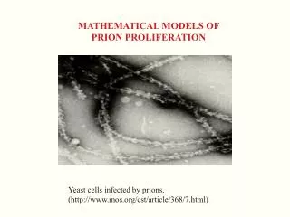
MATHEMATICAL MODELS OF PRION PROLIFERATION. Yeast cells infected by prions. (http://www.mos.org/cst/article/368/7.html). Prion Proliferation Models Research Team. Meredith Greer (Bates College, Lewiston, Maine, USA) Hans Engler (Georgetown University, Washington, DC, USA)

E N D
MATHEMATICAL MODELS OF PRION PROLIFERATION Yeast cells infected by prions. (http://www.mos.org/cst/article/368/7.html)
Prion Proliferation Models Research Team Meredith Greer (Bates College, Lewiston, Maine, USA) Hans Engler (Georgetown University, Washington, DC, USA) Jan Pruss (Martin Luther Universitat, Halle-Wittenberg, Germany) Laurent Pujo-Menjouet (University of Lyon, Lyon, France) Gieri Simonett (Vanderbilt University, Nashville, Tennessee, USA) Christoph Walker (Vanderbilt University, Nashville, Tennessee, USA) Glenn Webb (Vanderbilt University, Nashville, Tennessee, USA) Rico Zacher (Martin Luther Universitat, Halle-Wittenberg, Germany)
Transmissible Spongiform Encephalopathies (TSEs) TSEs are diseases such as Creutzfeld-Jakob disease in humans, scrapie in sheep, and bovine spongiform encephalopathies in cows. These diseases are characterized by long incubation periods, lack of immune response, and invisibility to detection as viruses. In 1982 Stanley Prusiner postulated that these diseases are caused not by viruses, but by abnormally shaped proteins, which he called prions. This hypothesis explains many of the features of the infectious agents of TSEs, except for their ability to replicate. Prions lack DNA or RNA , which is the commonly accepted basis for replication. Current research in this field seeks to explain the mechanism of prion replication.
The nucleated polymerization theory J. Jarrett and P. Lansbury, Cell, 1993 M. Eigen, Biophys. Chem, 1996 The leading theory of prion replication is nucleated polymerization. We use the notations for the normal PrPC (prion protein cellular) and abnormal PrPSc (prion protein scrapies) to denote the two primary forms of prions. By polymerize we mean that PrPSc increases its length by attaching units of PrPC in a string-like fashion. After a monomer attaches to the polymer, it is converted to the infectious form. Once the PrPSc is long enough to wrap into a helical shape (the nucleus), it forms stabilizing bonds that constitute the polymer strings. These strings can be formed into lengths of thousands of monomer units.
Replication of prion polymers by splitting PrPSc polymers may split into two smaller polymers, which results in two infectious polymers capable of further lengthening. If after splitting, a smaller polymer falls below the critical size, however, it degrades immediately into normal PrPC monomers. The biological processes are (1) lengthening (by addition of PrPC monomers), splitting (into two smaller polymer lengths), and degradation (by metabolic processes)
An infinite system of ODE model J. Masel, V. Jansen, M. Nowak, Biophys. Chem. 1999
A model with continuous polymer length V(t) = population of normal PrPC monomers at time t u(x,t) = density of polymers at time t w.r.t. length x in (x0, ), (where x0 > 0 is the minimum length) Let U(t) = total polymer population at timet. U(t)
Dynamics of the monomer population l = background source of monomers g = degradation rate of monomers t= conversion rate of monomers to polymers b(y) = rate of splitting of monomers to polymers k(x,y) = probability that a polymer of length y splits to lengths x and y-x
Steady states for the associated system of ODEs The disease free steady state: The disease steady state:
Linearization about the disease-free steady state The linearization about the disease free steady state V = l/g, U = 0, P = 0 is The eigenvalues are Theorem. The steady state V = l/g, U = 0, P = 0is locally exponentially asymptotically stable if
Linearization about the disease steady state which exists in the positive cone if
The inearization about the disease steady state is The eigenvalues of the linearization satisfy the characteristic equation where
By the Ruth-Hurwitz condition the eigenvalues all have negative real parts iff Theorem. The prion disease steady state is locally exponentially asymptotically stable if
Lyapunov functionals If s<x r, then a Lyapunov functional for disease free equilibruim is If s > x r, then a Lyapunov functional for disease equilibruim is
Convergence to the disease steady state The parameters are taken from J. Masel, V. Jansen, M. Nowak, Biophys. Chem. 1999 and R. Rubenstein et al., J. Infect. Dis. 1991.l=4400 , t=0.3, g=5.0, b=.0001, m=0.04, x0=6
Phase portrait of V(t), U(t), and P(t) All solutions converge to the disease steady state (V,U,P) = (55, 250, 103,132).
Application to a model of HIV infection D. Ho et al., Rapid turnover of plasma virions and CD 4 lymphocytes in HIV-1 infection, Nature 1995, and M. Nowak and R. May, Virus Dynamics, 2000 I(t) = infected CD4+ T cells at time t T(t) = uninfected CD4+ T cells at time t V(t) = virus at time t
Asymptotic behavior of the model of HIV infection Let R0 = b l N / d g. R0 is the number of secondary infections produced by one infectious virus in a totally susceptible population. If R0 < 1, then all solutions converge to the disease free steady state Tss =l/d, Iss = 0, Vss = 0. If R0 > 1, then all solutions converge to the disease steady state
The case R0 < 1 Let d = .01, l = 10, b = 10-4.1, N = 250, m = .5, g = 24. R0 = .827. All solutions converge to the disease free steady state Tss=1000, Iss = 0, Vss = 0.
The case R0 > 1 Let d = .01, l = 10, b = 10-4.1, N = 1000, m = .5, g = 24. R0 = 3.31. All solutions converge to the disease steady state Tss =302, Iss = 14, Vss = 291.
Application to an SEIS epidemic model S(t) = susceptible population at time t E(t) = exposed population at time t (infected but not yet infectious) I(t) = infectious population at time t
Asymptotic behavior of the SEIS epidemic model Let R0 = a l t / [g (a +m) (b+n)]. R0 is the number of secondary infections produced by one infective in a totally uninfected susceptible population. If R0 < 1, then all solutions converge to the disease free steady state Sss =l/g, Ess = 0, Iss = 0. If R0 > 1, then all solutions converge to the disease steady state
The case R0 < 1 Let b = 0.05, l = 500, m =0.1, t=10-6.9, a =0 .2, g = .0003, n=.1. R0 = .933. All solutions converge to the disease free steady state Sss =1,666,667, Ess = 0, Iss =0.
The case R0 > 1 Let b = .05, l = 500, m =.1, t=10-6.5, a = .2, g = .0003, n=.1. R0 = 2.34. All solutions converge to the endemic steady state Sss =711,512, Ess = 1228, Iss = 1637.
Ideas of the proof The solution V(t) can be considered known. Let w(t) = tV(t). w(t) converges exponentially to w*= l/g in the disease free case and to w*=(m + b x0)2/bt in the disease case. First consider the autonomous equation for u(x,t), where w(t) = w*. Prove that that there is a strongly continuous, linear, positive, contraction (exponentially in the disease free case) semigroup e-t L, t> 0 in the space X = L1((x0,);x dx) associated with the autonomous equation. Prove that the resolvent of L is compact in X, and thus has only point spectrum in the closed right-half plane. Show that 0 is the only eigenvalue of L on the imaginary axis, it is simple, the ergodic projection P onto the kernel on N(L) of L along the range R(L) of L exists and is rank one, find a formula for P, and prove that e-t L converges strongly to P in X. Use the method of characteristics to prove that the nonautonomous equation for u(x,t) is well-posed, obtain bounds for ux(.,t) in X, and use the convergence of w(t) to w*toshow that u(.,t) converges in X to the equililbrium u*.
Model simulation compared to experimental data of scrapie associated fibril counts SAF measurements after intracerebral injection of the 139A scrapie strain into Compton white mice from Rubenstein et al., J. Infect. Dis., 1991.The arrow indicates the onset of symptoms. The parameters of the simulation arel=4400 , t=0.3, g=5.0, b=.0001, m=0.04, x0=6.
Evolution of the polymer density u(x,t) The polymer density u(x,t) converges to the steady state.
Evolution of the mean length P(t)/U(t) of the polymer density The length of the average polymer u(x,t) increases fast initially and then slowly decreases due to the depletion of the PrPC monomer population.
Linear dependence on the log scale of the incubation times vs the inoculum dose The linear dependence of the incubation times on the log scale for nine orders of magnitude of inoculum dilutions.
More general models Allow the parameters m and b to depend on polymer length x. Allow the splitting kernel k(x,y) to have a more general form
References H. Engler, J. Pruss, and G. Webb, Analysis of a model for the dynamics of prions II, to appear in J. Math. Anal. Appl. M. Greer, L. Pujo-Menjouet, and G. Webb, A mathematical analysis of the dynamics of prion proliferation, to appear. J. Masel, V. Jansen, and M. Nowak, Quantifying the kinetic parameters of prion replication, Biophysical Chemistry 77 (1999) 139-152. Nowak, M., et al. Prion infection dynamics, Integrative Biology 1 (1998) 3-15. Prusiner, S. Molecular biology of prion diseases, Science 252 (1991) 1515-1522. J. Pruss, L. Pujo-Menjouet, G. Webb, and R. Zacher, Analysis of a model for the dynamics of prions, to appear in Discr. Cont. Dyn. Sys. Rubenstein, R. et al., Scrapie-infected spleens: analysis of infectivity, scrapie-associated fibrils, and protease-resistant proteins, J. Infect. Dis. 164, (1999) 29-35. Simonett, G. and Walker, C., On the solvability of a mathematical model of prion proliferation, to appear.