Download
1 / 28
300 likes | 827 Views
INFRARED SPECTROSCOPY. Pramod K Singh School of Basic Sciences &Research Sharda University, Greater Noida, INDIA. What is Infrared ?. IR radiation lies betn the visible and microwave portions of the em spectrum. *Infrared waves have wavelengths longer than visible and shorter

E N D
INFRARED SPECTROSCOPY Pramod K Singh School of Basic Sciences &Research Sharda University,Greater Noida, INDIA
What is Infrared? IR radiation lies betn the visible and microwave portions of the em spectrum. *Infrared waves have wavelengths longer than visible and shorter than microwaves, and have frequencies which are lower than visible and higher than microwaves. • The IR region is divided into: near, mid and far-infrared. • Near-infrared refers to the part of the infrared spectrum that is closest to visible light • far-infrared refers to the part that is closer to the microwave region. • Mid-infrared is the region between these two.
What is Infrared? • The primary source of infrared radiation is thermal radiation. (heat) • It is the radiation produced by the motion of atoms and molecules in an object. • The higher the temperature, the more the atoms and molecules move and the more infrared radiation they produce. • Any object radiates in the infrared. Even an ice cube, emits infrared.
*The IR region of the em spectrum covers a wide range of wavelengths, from about 800 nm (end of the visible region) to about 0.2 mm (beginning of the microwave region). *The units of wavelength commonly employed in the infrared region is wave number, = 1/, in reciprocal cm (cm-1).
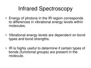
Infrared radiations • IR does not have sufficient energy to cause the excitation of e, however it causes atoms and group of atoms of organic compounds to vibrate faster about the covalent bonds which connect them. • The vibrations are quantised as they occur, the compound absorbs infrared energy in particular regions of the spectrum.
What is a vibration in a molecule? Any change in shape of the molecule- stretching of bonds, bending of bonds, or internal rotation around single bonds
Molecular Vibrations Covalent bonds vibrate at only certain allowable frequencies.
Calculation of vibrational frequencies :Let us consider a chemical bond to be a stretched spring. Then according to the Hooke’s law the frequency of vibration depends upon the strength of the bond. Stronger the bond more is the amount of energy required to stretch it. Wave numbers are proportional to energy. K is force constant
Conditions for I R activity (Selection Rule) The necessary conditions for a molecule to be IR active are: *The frequency of IR radiation must be equal to the vibrating frequency of a particular group or bond in the molecule *The molecule must have finite dipole moment. * If it does not have dipole moment,Some of its vibrations must produce an induced dipole moment.
Asymmetrical stretching/bending and internal rotation change the dipole moment of a molecule. Asymmetrical stretching/bending are IR active. Symmetrical stretching/bending does not and hence Not IR active
IR-Active and Inactive • A polar bond is usually IR-active. • A nonpolar bond in a symmetrical molecule will absorb weakly or not at all.
Infrared spectrometer double – beam infrared spectrometer. Its essential features are described briefly as follows:
Radiation source : • *Small ceramic rod, heated electrically in the range 1100 – 18000C and is made of either silicon carbide (Glowbar) or Nernst filament (a high – resistance, brittle element composed of mixture of sintered oxides of zirconium, thorium and cerium held together by a binding material). • The radiation is divided into two beams, one of which passes through the sample while the other function as a reference beam. The reference and the sample beams are then passed alternately into a monochromator at very short intervals by means of a rotating mirror.
(ii) Absorption cells and sample preparation : The cells generally employed are made up of rock salt or potassium bromide *(glass and quartz cells are unsuitable as these materials themselves absorb IR radiation).
(iii) Monochromator : The pulse beam enters the monochromator through an entrance slit and is dispersed by a grating or by a Littrowmount prism. In the monochromator the emergent beams are sorted out into individual wave lengths by means of sodium chloride prism which is transparent to infrared radiation throughout the range 4000 – 550 cm-1.
(iv) Detector, Amplifier and Recorder : The pulsating single beam now emerging through the exit slit is a narrow band consisting of only a very few frequencies After this sorting out (dispersion), the beams are focused alternately at each particular wave length throughout the spectral range, by means of a mirror system on to the detector, usually a sensitive fast thermocouple.
The signals from this are amplified electronically and by means of a mechanical arrangement, the spectrum is recorded on a special graph paper mounted on a rotating drum.
FT-IR Spectrometer • Fourier transform infrared spectroscopy is a technique for obtaining high quality infrared spectra by mathematical conversion of an interference pattern into a spectrum. • Uses an interferometer. • Has better sensitivity. • Less energy is needed from source. • Completes a scan in 1-2 seconds. • Takes several scans and averages them. • Has a laser beam that keeps the instrument accurately calibrated.
FT Optical System Diagram FTIR seminar Light source He-Ne gas laser (ceramic) Beam splitter Movable mirror Sample chamber (DLATGS) Fixed mirror Detector Interferometer
How the IR spectra of solid, liquid and gaseous compounds recorded?1. The spectrum of a solid sample is determined with an alkali halide pellet. About 1-3 mg of substance and 100-200 mg of alkali halide are ground together, dried to remove moisture and pressed at room temperature under high pressure into a small disc. KBr does not absorb IR radiation in the region 4000-650 cm-1 a complete spectrum of the solid is obtained.2. For free flowing liquids a neat spectrum may be recorded.3. Spectrum of a substance in a solution can also be done by using cells of 0.1 mm with 10% solution are used. CCl4, CS2 and CHCl3 are the solvents to be used.4. The spectra of gases or low boiling liquids can be obtained by expansion of the sample into an evacuated cell.
Carbonyl Stretching • The C=O bond of simple ketones, aldehydes, and carboxylic acids absorb around 1710 cm-1. • Usually, it’s the strongest IR signal. • Carboxylic acids will have O-H also. • Aldehydes have two C-H signals around 2700 and 2800 cm-1.
O-H Stretch of a Carboxylic Acid This O-H absorbs broadly, 2500-3500 cm-1, due to strong hydrogen bonding.