Download
1 / 72
820 likes | 1.29k Views
Chapter 8. Cytoskeleton System. Biology Department of the Basic Teaching Colledge. Xiamixinuer · Yilike. March of 2012. Teaching Requirements:. 1. Mastering: concepts of the cytoskeleton; structure, chemical composition, and assembly of microtubules and microfilaments.

E N D
Chapter 8 CytoskeletonSystem Biology Department of the Basic Teaching Colledge Xiamixinuer·Yilike March of 2012
Teaching Requirements: 1. Mastering: concepts of the cytoskeleton; structure, chemical composition, and assembly of microtubules and microfilaments. 2. Comprehending: functions of microtubules and microfilaments. 3. Understanding: functions of the cytoskeleton; types and functions of intermediate filaments.
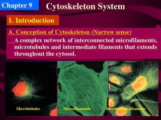
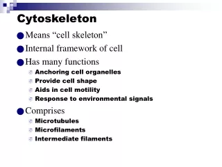
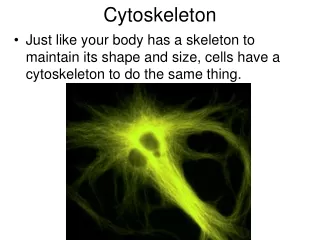
1. Introduction A. Conception of Cytoskeleton (Narrow sense) A complex network of interconnected microfilaments, microtubules and intermediate filaments that extends throughout the cytosol.
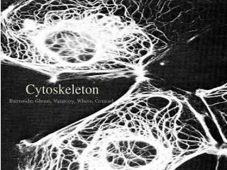
Fluorescent microscopy and Electron microscopy : • Immunofluorescence: fluorescently-labeled antibody • Fluorescence: microinject into living cells • Video microscopy: in vitro motility assays • Electron: Triton X-100, Metal replica • Drugs and mutations (about functions) • Biochemical analysis(in vitro) B. Techniques for studying the cytoskeleton
Fluorescence microscopy microtubules actin filamin cytoskeleton microfilaments microtubules microtubules
C. The self-assembly and dynamic structure of cytoskeletal filaments • Each type of cytoskeletal filament is constructed from smaller protein subunits. • The cytoskeleton is a network of three filamentous structures. • The cytoskeleton is a dynamic structure with many roles.
D. The function of the cytoskeleton • Structural support • Internal framework maintaining position of the organelles • Machinery required for movement of materials and organelles within cells • Force generating elements responsible for movement of cells from one place to another
2.Microtubule, MT A. Structures: • Hollow • Tubular structures 25nm in diameter • Assembled from protein tubulin • The tubulin consists of alpha-beta tubulin heterodimers arranged in rows (protofilaments) • Form cytoskeleton, mitotic spindle, centrioles, core of cilia and flagella
a and ßTubulin heterodimers are the protein building blocks of MTs
Arrangement of protofilaments in singlet, doublet, and triplet MTs Singlet Doublet Triplet A A B B C In cilia and flagella In centrioles and basal bodies
Assembling process of MT OUTSIDE OF THE BODY + PEDAL -
assemble Head tail connection 2 3 1 4 13 5 12 6 11 7 10 9 8 CROSSSECTION tubulin heterodimer MT (13) profilament tubulin
B. MTs assemble from microtubule-organizing centers (MTOCs) Microtubule-organizing centers (MTOCs):is the region to assemble MT,Where includes-tubulin. MTOCs:includeCentrosome, Mitotic spindle and Basal body.
(1) Interphase: Centrosome Microtubule-organizing centers (MTOCs) Dynamic instability (2) Dividing cell: Mitotic spindle Dynamic instability (3) Ciliated cell: Basal body Stability
Centrioles Centrioles are short cylinders with a 9 + 0 pattern of microtubule triplets. Centrioles may be involved in microtubule formation and disassembly during cell division and in the organization of cilia and flagella.
MT are nucleated by a protein complex containing -tubulin The centrosome is the major MTOC of animal cells
Why the centrosome can act as MTOC? Treat cell with colcemid Cytosolic MTs depoly, except those in centrosome A • Experiments supporting that centrosome is the MTOC Expla I: MTOC nucleate poly of tubulins Remove colcemid Tublin repoly Expla II: MTOC gather MTs in cytosol B centrosome + Tubulins MT + Tubulins No
Cilia and flagella Cilia (small and numerous) and flagella (large and single) have a 9 + 2 pattern of microtubules and are involved in cell movement. Cilia and flagella move when the microtubule doublets slide past one another. Each cilium and flagellum has a basalbody at its base.
Dynamic instability due to the structural differences between a growing and a shrinking microtubule end. • GTP cap; • Catastrophe: accidental loss of GTP cap; • Rescue: regain of GTP cap C. Characteristics of MT assembly
Microtubules have a plus and minus ends. • Typically the minus is for anchoring and the plus is for growing. • The transition between MT growth and MT shrinking is controlled in cells by special proteins..
Drugs affect the assembly of MTs (1) Colchicine Binding to tubulin dimers, prevent MTs polymerization (2)Taxol Binding to MTs, stabilize MTs These compounds are called antimitotic drugs, and have application in medical practice as anticancer drugs
MAPs modulate MT structure, assembly, and function D. Microtuble-associated proteins (MAPs) Control organization Katanin like proteins MAPs Tau: In axon, cause MTs to form tight bundles MAP2: In dendrites, cause MTs to form looser bundles MAP1B: In both axons and dendrites to form crossbridge between microtubules
5. Functions of MTs • A. Maintenance of cell shape(constitute the centriols and cilia or flagella). • B. Cell motility (see in cilia or flagella). • C. Chromosome movements in cell division • D. Organelle movement (MT associated motor proteins: kinesins: towards + end (anterograde transport) Golgi to ER or PM traffic;dyneins: towards - end (retrograde transport) ER to Golgi traffic.)
5. Functions of MTs A. Maintenance of cell shape(constitute the centriols and cilia or flagella).
Constitute the centriols and cilia or flagella • No centrioles in Plant and fungi • A pair of centrioles are surrounded by electron dense pericentriolar material. • Centrioles contain nine evenly spaced fibrils, each containing three microtubules, A, B and C tubules. • A tubule is connected to the center of the centriole by a radial spoke. • Centrioles are in pairs and at right angles to each other. Structure
5. Functions of MTs • B. Cell motility (see in cilia or flagella).
Motility of MT(CILIA,FILAGELA MOVEMENT) SPERM MOVEMENT CILIA MOVEMENT
Dyenin arms responsible for sliding Crosslinks and spokes responsible for bending
B. Transport in the cytoplasm MT associated motor proteins: kinesins: towards + end (anterograde transport) Golgi to ER or PM traffic dyneins: towards - end (retrograde transport) ER to Golgi traffic
3. Microfilament, MF • Using ATP, G-actin polymerizes to form MF(F-actin) A. MFs are made of actin and involved in cell motility.
MINUS END F-actin G-actin PLUS END
Assembly of MF -end G-actin Dimer Trimer +end F-actin
Characteristics: (1) Within a MF, all the actin monomers are oriented in the same direction, so MF has a polarity B. MF assembly and disassembly Myosin is molecular motor for actins.
(2) In vitro, (Polymerization) both ends of the MF grow, but the plus end faster than the minus. Because actin monomers tend to add to a filament’s plus end and leave from its minus end---- “Tread-milling”
(3) Dynamic equilibrium between the G-actin and polymeric forms, which is regulated by ATP hydrolysis and G-actin concentration.
(4) Dynamic equilibrium is required for the cell functions. Some MFs aretemporary and others permanent.