Download
1 / 52
520 likes | 527 Views
Chapter 4 Physiology of Cells. Two Ways Molecules Move through Cells. 1) Passive Transport 2) Active Transport. Movement of Substances through Cell Membranes. Passive transport processes—do not require any energy expenditure of the cell membrane 4 types of passive transport 1) diffusion

E N D
Two Ways Molecules Move through Cells • 1) Passive Transport • 2) Active Transport
Movement of Substances through Cell Membranes • Passive transport processes—do not require any energy expenditure of the cell membrane • 4 types of passive transport • 1) diffusion • 2) Dialysis • 3) Osmosis • 4) Facilitated diffusion
Diffusion • Diffusion—a passive process • Molecules spread through the membranes • Molecules move from an area of high concentration to an area of low concentration, down a concentration gradient • As molecules diffuse, a state of equilibrium will occur
Simple diffusion • Simple diffusion • Molecules cross through the phospholipid bilayer • Solutes permeate the membrane; therefore, we call the membrane permeable
Osmosis • Osmosis • Diffusion of water through a selectively permeable membrane, which limits the diffusion of at least some of the solute particles • Water pressure that develops as a result of osmosis is called osmotic pressure
Osmosis Cont. • Potential osmotic pressure is the maximum pressure that could develop in a solution when it is separated from pure water by a selectively permeable membrane; knowledge of potential osmotic pressure allows the prediction of the direction of osmosis and the resulting change of pressure
Osmosis • Isotonic—two fluids that have the same potential osmotic pressure
Osmosis • Hypertonic—“higher pressure”; cells placed in solutions that are hypertonic to intracellular fluid always shrivel as water flows out of cell; this has great medical importance: if medical treatment causes the extracellular fluid to become hypertonic to the cells of the body, serious damage may occur
Osmosis • Hypotonic—“lower pressure”; cells placed in a hypotonic solution may swell as water flows into them; water always osmoses from the hypotonic solution to the hypertonic solution
Facilitated diffusion • Facilitated diffusion (mediated passive transport) • A special kind of diffusion whereby movement of molecules is made more efficient by the action of transporters embedded in a cell membrane • Transports substances down a concentration gradient • Energy required comes from the collision energy of the solute
Facilitated diffusion • Channel-mediated passive transport • Channels are specific—allow only one type of solute to pass through • Gated channels may be open or closed (or inactive)—may be triggered by any of a variety of stimuli • Channels allow membranes to be selectively permeable • Carriers attract and bind to the solute, change shape, and release the solute out the other side of the carrier • Carriers are usually reversible, depending on the direction of the concentration gradient
Active Transport • Active transport is the movement of solute particles from an area of low concentration to an area of high concentration (up the concentration gradient) by means of a carrier molecule. • There are three types: • 1) Phagocytosis (Endocytosis) • 2) Pinocytosis (Endocytosis) • 3) Exocytosis
Endocytosis • Endocytosis—the plasma membrane “traps” some extracellular material and brings it into the cell in a vesicle • Two basic types of endocytosis: • Phagocytosis—“condition of cell-eating”; large particles are engulfed by the plasma membrane and enter the cell in vesicles; vesicles fuse with lysosomes, where the particles are digested • Pinocytosis—“condition of cell-drinking”; fluid and the substances dissolved in it enter the cell
Exocytosis • Exocytosis • Process by which large molecules, notably proteins, can leave the cell even though they are too large to move out through the plasma membrane • Large molecules are enclosed in membranous vesicles that are then pulled by the cytoskeleton to the plasma membrane, where the contents are released • Exocytosis also provides a way for new material to be added to the plasma membrane
Cell Metabolism • Metabolism is the set of chemical reactions in a cell • Catabolism—breaks large molecules into smaller ones; usually releases energy • Anabolism—builds large molecules from smaller ones; usually consumes energy
Enzymes • Role of enzymes • Enzymes are chemical catalysts, reducing activation energy needed for a reaction • Enzymes regulate cell metabolism • Chemical structure of enzymes • Proteins of a complex shape • The active site is where the enzyme molecule fits the substrate molecule—the lock-and-key model
6.3 Section Summary 6.3 – pages 157-163 The structure of proteins • Enzymes are important proteins found in living things. An enzyme is a protein that changes the rate of a chemical reaction. • They speed the reactions in digestion of food. • YouTube - Enzyme
Classification and naming of enzymes • Enzymes usually have an -ase ending, with the first part of the word signifying the substrate or the type of reaction catalyzed • Oxidation-reduction enzymes—known as oxidases, hydrogenases, and dehydrogenases; energy release depends on these enzymes • Hydrolyzing enzymes—hydrolases; digestive enzymes belong to this group
Classification and naming of enzymes (cont.) • Phosphorylating enzymes—phosphorylases or phosphatases; add or remove phosphate groups • Enzymes that add or remove carbon dioxide— carboxylases or decarboxylases • Enzymes that rearrange atoms within a molecule—mutases or isomerases • Hydrases add water to a molecule without splitting it
General functions of enzymes • Various chemical and physical agents known as allosteric effectors affect enzyme action by changing the shape of the enzyme molecule; examples of allosteric effectors include the following: • Temperature • Hydrogen ion (H+) concentration (pH) • Ionizing radiation • Cofactors • End products of certain metabolic pathways
General functions of enzymes (cont.) • Most enzymes catalyze a chemical reaction in both directions • Enzymes are continually being destroyed and are continually being replaced • Many enzymes are first synthesized as inactive proenzymes
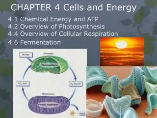
Catabolism • Cellular respiration, the pathway in which glucose is broken down to yield its stored energy, is an important example of cell catabolism • Cellular respiration has three pathways that are chemically linked: 1) Glycolysis 2) Citric acid cycle 3) Electron transport system (ETS)
Glycolysis (net yeild of 2 ATP) • Pathway in which glucose is broken apart into two pyruvic acid molecules to yield a small amount of energy (which is transferred to ATP and NADH) • Includes many chemical steps (reactions that follow one another), each regulated by specific enzymes • Is anaerobic (requires no oxygen) • Occurs within cytosol (outside the mitochondria)
Citric acid cycle (Krebs cycle) (net yield of 2 ATP • Pyruvic acid (from glycolysis) is converted into acetyl CoA and enters the citric acid cycle after losing CO2 and transferring some energy to NADH • Citric acid cycle is a repeating (cyclic) sequence of reactions that occurs inside the inner chamber of a mitochondrion. Acetyl splits from CoA and is broken down, yielding waste CO2 and energy (in the form of energized electrons), which is transferred to ATP, NADH, and FADH2
Electron transport system (ETS)(Net yield of 32 ATP) • Energized electrons are carried by NADH and FADH2 from glycolysis and the citric acid cycle to electron acceptors embedded in the cristae of the mitochondrion • As electrons are shuttled along a chain of electron-accepting molecules in the cristae, their energy is used to pump accompanying protons (H+) into the space between mitochondrial membranes
Electron transport system (cont.) • Protons flow back into the inner chamber through pump molecules in the cristae, and their energy of movement is transferred to ATP • Low-energy electrons coming off the ETS bind to oxygen and rejoin their protons, forming water (H2O)
Cell Metabolism • Anabolism • Protein synthesis is a central anabolic pathway in cells
Anabolism • Deoxyribonucleic acid (DNA) • A double-helix polymer (composed of nucleotides) that functions to transfer information, encoded in genes, that directs the synthesis of proteins • Gene—a segment of a DNA molecule that consists of approximately 1000 pairs of nucleotides and contains the code for synthesizing one polypeptide
Anabolism • Transcription—mRNA forms along a segment of one strand of DNA • Editing • Noncoding introns are removed, and remaining exons are spliced together to form the final, edited version of the mRNA copy of the DNA segment • Spliceosomes are ribosome-sized structures in the nucleus that splice mRNA transcripts
Anabolism (cont.) • Translation • After leaving the nucleus and being edited, mRNA associates with a ribosome in the cytoplasm • tRNA molecules bring specific amino acids to the mRNA at the ribosome; the type of amino acid is determined by the fit of a specific tRNA’s anticodon with mRNA’s codon • As amino acids are brought into place, peptide bonds join them—eventually producing an entire polypeptide chain
Growth and Reproduction of Cells • Cell growth and reproduction of cells are the most fundamental of all living functions and together constitute the cell life cycle • Cell growth—depends on using genetic information in DNA to make the structural and functional proteins needed for cell survival • Cell reproduction—ensures that genetic information is passed from one generation to the next
Growth and Reproduction of Cells • Cell growth—a newly formed cell produces a variety of molecules and other structures necessary for growth using the information contained in the genes of DNA molecules; this stage is known as interphase • Production of cytoplasm—more cell material is made, including growth and/or replication of organelles and plasma membrane; a largely anabolic process
Growth and Reproduction of Cells • Cell growth (cont.) • DNA replication (Table 4-4) • Replication of the genome prepares the cell for reproduction; mechanics are similar to RNA synthesis • DNA replication (Figure 4-29) • DNA strand uncoils and strands come apart • Along each separate strand, a complementary strand forms • The two new strands are called chromatids, instead of chromosomes • Chromatids are attached in pairs, and the centromere is the name of their point of attachment
Growth and Reproduction of Cells • Cell growth (cont.) • Growth phase of the cell life cycle can be subdivided into the first growth phase (G1), the [DNA] synthesis phase (S), and the second growth phase (G2)
Growth and Reproduction of Cells • Cell reproduction—cells reproduce by splitting themselves into two smaller daughter cells (Table 4-5) • Mitotic cell division—the process of organizing and distributing nuclear DNA during cell division has four distinct phases (Figure 4-31)
Growth and Reproduction of Cells • Mitotic cell division (cont.) • Prophase—“before-phase” • After the cell has prepared for reproduction during interphase, the nuclear envelope falls apart as the chromatids coil up to form chromosomes, which are joined at the centromere (Figure 4-30) • As chromosomes are forming, the centriole pairs are seen to move toward the poles of the parent cell and spindle fibers are constructed between them
Growth and Reproduction of Cells • Mitotic cell division (cont.) • Metaphase—“position-changing phase” • Chromosomes move so that one chromatid of each chromosome faces its respective pole • Each chromatid attaches to a spindle fiber
Growth and Reproduction of Cells • Mitotic cell division (cont.) • Anaphase—“apart phase” • Centromere of each chromosome has split to form two chromosomes, each consisting of a single DNA molecule • Each chromosome is pulled toward the nearest pole, forming two separate but identical pools of genetic information
Growth and Reproduction of Cells • Mitotic cell division (cont.) • Telophase—“end phase” • DNA returns to its original form and location within the cell • After completion of telophase, each daughter cell begins interphase to develop into a mature cell