Download
1 / 75
780 likes | 809 Views
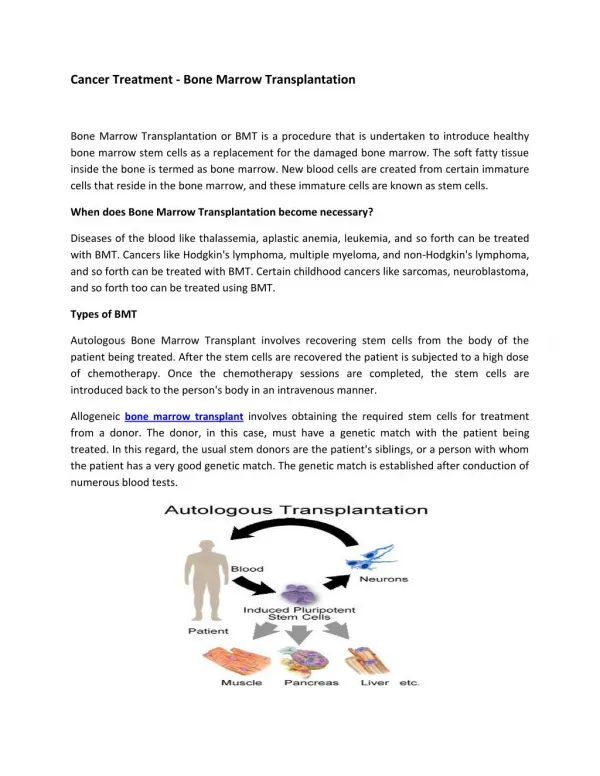
Patophysiology of blood and bone marrow disorders. Pathophysiology of blood. Patophysiology of RBC Basic information Hemoglobin Laboratory tests Iron Erythropoetin a erythropoesis Anemias Principles of light microscopy Observation of blood smears. Theoretical part. Practical part.

E N D
Pathophysiology of blood • Patophysiology of RBC • Basic information • Hemoglobin • Laboratory tests • Iron • Erythropoetin a erythropoesis • Anemias • Principles of light microscopy • Observation of blood smears Theoretical part Practical part
Definition of hematology conception Hematology(gr. haima-haimatos blood, gr. logos science- hematology, science about blood a blood disorders) is deal with blood and haemoplastic bodies • Peripheral blood • Red bone marrow • Lymph - nodes • Liver, spleen
Basic anatomical and physiological notes • Basic haemoplastic bodies: • Bone marrow • Thymus • Lymph-nodes • MALT • (mucosa associated lymphoid tissue) • Spleen • Peripheral blood Central haemoplastic and immune bodies Peripheral haemoplastic and immune bodies
Ontogenesis of haemopoiesis Yolk sack Liver: 6. week - birth Spleen, thymus, nodes: 8.- 16. week Red bone marrow: 12. week – Extramedullar haemopoiesis
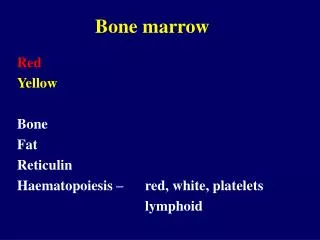
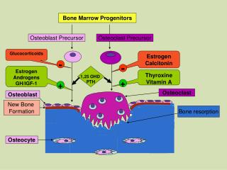
Bone marrow • Place of haemopoiesis • Red bone marrow • haemopoetic • Yellow bone marrow • Adiposal tissue
Red bone marrow • stroma • system of reticular cells • system of reticular fibres • collagen fibers • fibronectin, laminin, hemonectin • hematogenous filaments • sinusoidal capillaries
Factors necessary for haemopoiesis • Pluripotent stem cell • is able either mitotic or meiotic cell division • „Microenvironment“ – e.g. bone marrow • part of b.m are cells and ECM • Growth factors • so-called colony stimulating factors = CSF (are secreted glycoproteins which bind to receptor proteins on the surfaces of hemopoietic stem cells and thereby activate intracellular signaling pathways which can cause the cells to proliferate and differentiate into a specific kind of blood cell) • erythropoietin
SPECIFIC IMMUNE DEFENSE GAS TRANSPORT HEMOSTASIS IMMUNE RESPONSE
Structure of RBC • The mature red blood cell is easily recognized because of its unique morphology. • At rest, the red blood cell takes the shape of a biconcave disc with a mean diameter of 8 µm, a thickness of 2 µm, and a volume of 90 fL. • It lacks a nucleus or mitochondria, and 33% of its contents is made up of a single protein, hemoglobin. • Intracellular energy requirements are largely supplied by glucose metabolism, which is targeted at maintaining hemoglobin in a soluble, reduced state, providing appropriate amounts of 2,3-diphosphoglycerate (2,3-DPG), and generating adenosine triphosphate (ATP) to support membrane function. • Lifespan of RBC???
Red blood cell morphology A: Adult red blood cells are characterized by their lack of a nucleus, and biconcave disc shape. Red blood cells are extremely pliable as they pass through small vessels and sinusoids. B: The section of a small blood vessel demonstrates the ability of red blood cells to undergo major shape distortions.
Red Cell Production Committed bone marrow cells proliferate and differentiate through the erythroblast and normoblast stages to reticulocytes, which are released into the bloodstream and finally become erythrocytes.
Mature of RBC • erythropoetin, Fe, folate, vit. B12 • proerythroblast – lace chromatin • basophilic erythroblast – strong basophilic cytoplasma • due to synthesis of Hb • polychromatophilic erythroblast • orthochromatic erythroblast – no dividing • reticulocyte – expulsion of nucleus • rest of polyribosomes • the period from stem cell to emergence of the reticulocyte in the circulation normally takes approximately 1 week • maturation of reticulocyte to erythrocyte takes approximately 24 to 48 hours
Count of RBC Normal values:female: 3,8 – 5,2 x 10¹² / 1 litr male: 4,2 – 5,8 x 10¹² / 1 litr Decrease: anemia, expansion of ECF Increase: polycytemia, dehydratation
Destruction of red blood cells • The destruction of red blood cells is mediated by a group of large phagocytic cells found in the spleen, liver, bone marrow, and lymph nodes. • During red blood cell destruction, amino acids from the globin chains and iron from the heme units are salvaged and reused, whereas the bulk of the heme unit is converted to bilirubin, the pigment of bile.
Destruction of red blood cells • Bilirubin, which is insoluble in plasma, attaches to plasma proteins for transport to the liver, where it removed from the blood and conjugated with glucuronide to render it water soluble so that it can be excreted in the bile. • The plasma-insoluble form of bilirubin is referred to as unconjugated bilirubin the water-soluble form is referred to as conjugated bilirubin.
Hemoglobin • The red blood cell is, basically, a container for hemoglobin a 64,500 dalton protein made up of 4 polypeptide chains, each containing an active heme group. • Each heme group is capable of binding to an oxygen molecule. • The respiratory motion of hemoglobin, that is, the uptake and release of oxygen to tissues, involves a specific change in molecular structure
Cellular metabolism • The stability of the red blood cell membrane and the solubility of intracellular hemoglobin depend on four glucose-supported metabolic pathways: • EMBDEN-MEYERHOFF PATHWAY • METHEMOGLOBIN REDUCTASE PATHWAY • PHOSPHOGLUCONATE PATHWAY • LUEBERING-RAPAPORT PATHWAY
Red blood cell metabolic pathways The Embden-Meyerhoff pathway is responsible for the generation of high energy phosphate (ATP) for membrane maintenance, whereas the other pathways support hemoglobin function. The methemoglobin reductase (NADH-diaphorase) pathway is required to maintain hemoglobin in a reduced state. The phosphogluconate pathway helps counteract environmental oxidants. The Luebering-Rapaport pathway generates intracellular 2,3-DPG.
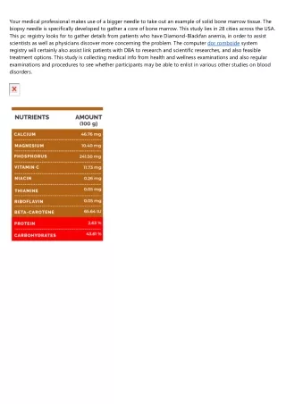
Iron metabolism • Body iron is found in several compartments. • About 80 % is complexed to heme in hemoglobin, and most of the remaining iron (about 20 %) is stored in the bone marrow, liver, spleen, and other organs. • Iron in the hemoglobin compartment is recycled. • When red blood cells age and are destroyed in the spleen, the iron from their hemoglobin is released into the circulation and returned to the bone marrow for incorporation into new red blood cells, or to the liver and other tissues for storage.
Iron metabolism • Dietary iron helps to maintain body stores. • Iron, principally derived from meat, is absorbed in the small intestine, especially the duodenum. Diagrammatic representation of the iron cycle, including its absorption from the gastrointestinal tract, transport in the circulation, storage in the liver, recycling from aged red cells destroyed in the spleen, and use in the bone marrow synthesis of red blood cells.
Iron metabolism • When body stores of iron are diminished or erythropoiesis is stimulated, absorption is increased. • Normally, some iron is sequestered in the intestinal epithelial cells and is lost in the feces as these cells slough off. • The iron that is absorbed enters the circulation, where it attaches a transport protein called transferrin. From the circulation, iron can be deposited in tissues such as the liver, where it is stored as ferritin , a protein–iron complex, which can easily return to the circulation. • Serum ferritin levels, which can be measured in the laboratory, provide an index of body iron stores.
Overview – red blood cell • The function of red blood cells, facilitated by the iron-containing hemoglobin molecule, is to transport oxygen from the lungs to the tissues. • The production of red blood cells, which is regulated by erythropoietin, occurs in the bone marrow and requires iron, vitamin B12, and folate. • The red blood cell, which has a life span of approximately 120 days, is broken down in the spleen; the degradation products such as iron and amino acids are recycled. • The heme molecule, which is released from the red blood cell during the degradation process, is converted to bilirubin and transported to the liver, where it is removed and rendered water soluble for elimination in the bile.
Laboratory tests • Red blood cells can be studied by means of a sample of blood. • In the laboratory, automated blood cell counters rapidly provide accurate measurements of red cell content and cell indices. • The (RBC) measures the total number of red blood cells in 1 mm3 of blood.
Laboratory tests • The percentages of reticulocytes (normally approximately 1%) provides an index of the rate of red cell production. • The hemoglobin (grams per dL of blood) measures the hemoglobin content of the blood. The major components of blood are the red cell mass and plasma volume.
Laboratory tests • The hematocrit measures the percentage of red cell mass in 100 mL of blood . • To determine the hematocrit, a sample of blood is placed in a glass tube, which is then centrifuged to separate the cells and the plasma. • The hematocrit may be deceptive because it varies with the quantity of extracellular fluid, rising with dehydration and falling with overexpansion of extracellular fluid volume.
Laboratory tests • Red cell indices are used to differentiate types of anemias by size or color of red cells: • The mean corpuscular volume (MCV) reflects the volume or size of the red cells • The mean corpuscular hemoglobin concentration (MCHC) is the concentration of hemoglobin in each cell - (anemias are described as normochromic (normal color or MCHC) or hypochromic (decreased color or MCHC). • Mean cell hemoglobin (MCH) refers to the mass of the red cell and is less useful in classifying anemia
Pathology of blood and haemoplastic functions • Lack of blood elements • Abundance of blood elements • Haematological malignits • Bleeding states • Trombotical states
Anemia – def. Anemia is defined as an abnormally low hemoglobin level, number of circulating red blood cells, or both, resulting in diminished oxygen-carrying capacity of the blood. • Anemia usually results from excessive loss (i.e., bleeding) or destruction (i.e., hemolysis) of red blood cells or from deficient red blood cell production because of a lack of nutritional elements or bone marrow failure.
Anemia • Anemia is not a disease, but an indication of some disease process or alteration in body function. The manifestations of anemia can be grouped into three categories: • those resulting from tissue hypoxia due to decreased oxygen delivery, • those due to compensatory mechanisms, and • the signs and symptoms associated with the pathologic process causing the anemia. • The manifestations of anemia depend on its severity, the rapidity of its development, and the affected person's age and health status.
Types of anemias • Blood loss anemia • Hemolytic anemias • Inherited disorders of the red cell membrane • Hemoglobinopathies • Sickle cell disease • Thalassemias • Inherited enzyme defects • Acquired hemolytic anemias • Anemias of deficient red cell production • Iron deficiency anemia • Megaloblastic anemia • Cobalamin-deficiency anemia • Folic acid-deficiency anemia • Aplastic anemia • Chronic disease anemia
Blood loss anemia • The clinical and red cell manifestations associated with blood loss anemia depend on the rate of hemorrhage and whether the bleeding loss is internal or external. • With rapid blood loss, circulatory shock and circulatory collapse may occur. • With more slowly developing anemia, the amount of red cell mass lost may reach 50% without the occurrence of signs and symptoms. • The effects of acute blood loss are mainly due to loss of intravascular volume, which can lead to cardiovascular collapse and shock.
Blood loss anemia • A fall in the red blood cell count, hematocrit, and hemoglobin is caused by hemodilution resulting from movement of fluid into the vascular compartment. Initially, the red cells are normal in size and color (normocytic, normochromic). • The hypoxia that results from blood loss stimulates proliferation of committed erythroid stem cells in the bone marrow. • It takes about 5 days for the progeny of stem cells to fully differentiate, an event that is marked by increased reticulocytes in the blood. • If the bleeding is controlled and sufficient iron stores are available, the red cell concentration returns to normal within 3 to 4 weeks. • External bleeding leads to iron loss and possible iron deficiency, which can hamper restoration of red cell counts.
Blood loss anemia • Chronic blood loss does not affect blood volume but instead leads to iron-deficiency anemia when iron stores are depleted. • Because of compensatory mechanisms, patients are commonly asymptomatic until the hemoglobin level is less than 8 g/dL. • The red cells that are produced have too little hemoglobin, giving rise to microcytic hypochromic anemia.
Red cell characteristics seen in different types of anemia (A) microcytic and hypochromic red cells, characteristic of iron-deficiency anemia; (B) macrocytic and misshaped red blood cells, characteristic of megaloblastic anemia; (C) abnormally shaped red blood cells seen in sickle cell disease; and (D) normocytic and normochromic red blood cells, as a comparison.
Hemolytic anemias • Hemolytic anemia is characterized by the premature destruction of red cells, retention in the body of iron and other products of hemoglobin destruction, and an increase in erythropoiesis to compensate for the loss of red cells. • Because of the red blood cell's shortened life span, the bone marrow usually is hyperactive, resulting in an increase in the number of reticulocytes in the circulating blood. • As with other types of anemias, the person experiences easy fatigability, dyspnea, and other signs and symptoms of impaired oxygen transport.
Hemolytic anemias • Hemolytic anemia can be further classified as to whether the underlying cause of the disorder is inherited or acquired. • Inherited disorders include the inherited disorders of the red cell membrane, hemoglobinopathies (i.e., sickle cell anemia and thalassemias), and inherited enzyme disorders. • Acquired forms of hemolytic anemia are caused by agents extrinsic to the red blood cell, such as drugs, bacterial and other toxins, antibodies, and physical trauma.
Hemoglobinopathies • The hemoglobinopathies represent disorders of the hemoglobin molecule, most being caused by point mutations in a globin chain gene. • The production of each type of globin chain is controlled by individual structural genes with five different gene loci. • Mutations can occur anywhere in these five loci. a) Sickle cell disease b) Thalassemia
1. Hereditary sideroblastic anemia 2. Sickle cell disease
Sickle cell disease • Sickle cell disease is an inherited disease that is caused by the presence of an abnormal hemoglobin S (HbS), which upon deoxygenation transforms the erythrocyte into a sickle shape. • HbS is transmitted by recessive inheritance and can manifest as sickle cell trait (i.e., heterozygote with one HbS gene) or sickle cell disease (i.e., homozygote with two HbS genes). • Sickle cell disease affects approximately 50,000 (0.1% to 0.2%) African Americans, and about 10% of African Americans carry the trait. • In parts of Africa, where malaria is endemic, the gene frequency approaches 30%, attributed to the slight protective effect it confers against Plasmodium falciparum malaria.