Download
1 / 2
20 likes | 133 Views
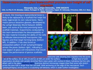
Nano-macro porous glass bone-scaffold provides new insight of cell migration Himanshu Jain, Lehigh University, DMR 0602975 USA. Co-PIs: R. M. Almeida, Lisbon, Portugal; M. Marei, Alexandria, Egypt; W. Soboyejo, Princeton, USA; A.C. Beye, Dakar, Senegal.

E N D
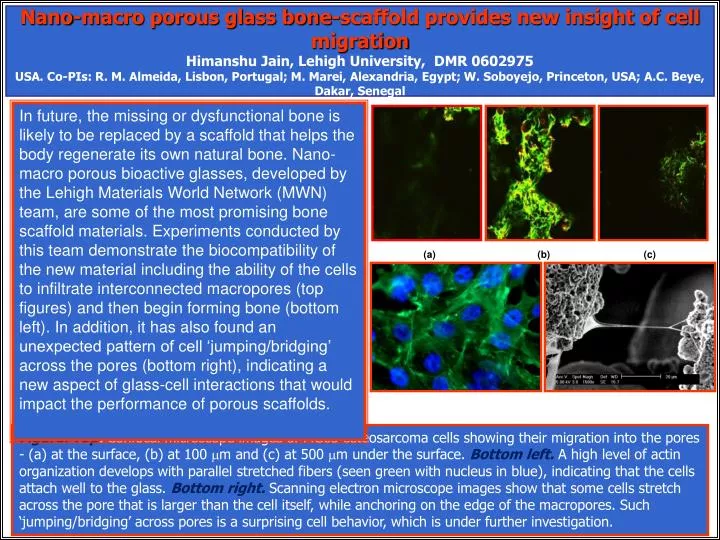
Nano-macro porous glass bone-scaffold provides new insight of cell migration Himanshu Jain, Lehigh University, DMR 0602975 USA. Co-PIs: R. M. Almeida, Lisbon, Portugal; M. Marei, Alexandria, Egypt; W. Soboyejo, Princeton, USA; A.C. Beye, Dakar, Senegal In future, the missing or dysfunctional bone is likely to be replaced by a scaffold that helps the body regenerate its own natural bone. Nano-macro porous bioactive glasses, developed by the Lehigh Materials World Network (MWN) team, are some of the most promising bone scaffold materials. Experiments conducted by this team demonstrate the biocompatibility of the new material including the ability of the cells to infiltrate interconnected macropores (top figures) and then begin forming bone (bottom left). In addition, it has also found an unexpected pattern of cell ‘jumping/bridging’ across the pores (bottom right), indicating a new aspect of glass-cell interactions that would impact the performance of porous scaffolds. (a) (b) (c) Figure. Top: Confocal microscope images of MG63 osteosarcoma cells showing their migration into the pores - (a) at the surface, (b) at 100 mm and (c) at 500 mm under the surface. Bottom left. A high level of actin organization develops with parallel stretched fibers (seen green with nucleus in blue), indicating that the cells attach well to the glass. Bottom right. Scanning electron microscope images show that some cells stretch across the pore that is larger than the cell itself, while anchoring on the edge of the macropores. Such ‘jumping/bridging’ across pores is a surprising cell behavior, which is under further investigation.
Pooled resources create new opportunity for multidisciplinary student training Himanshu Jain, Lehigh University, DMR 0602975 USA. Co-PIs: R. M. Almeida, Lisbon, Portugal; M. Marei, Alexandria, Egypt; W. Soboyejo, Princeton, USA; A.C. Beye, Dakar, Senegal The training of next generation of researchers with expertise both in biological and materials sciences remains a challenge. So Profs. Falk (Bio Sci) and Jain (Mat Sci) teamed up and leveraged the resources of this MWN project and the Int’l Materials Institute for New Functionality in Glass to win a grant from BSDI program funded by Howard Hughes Medical Institute. As a result, four undergraduate students are trained intensively by a team of 2 cell biologists, 3 glass scientists and a dentist. During the summer, these students split their time equally between the fabrication of nano-macro porous glass, and assessment of the proliferation of MG63 osteosarcoma and MC3T3 preosteoblast cells on their samples. Next, they plan to continue the investigation, more or less independently, in the following semester. Materials World Network Figure. Top: IMI-NFG visiting professor from Egypt, Dr. Moawad teaches glass fabrication to BDSI scholars. Bottom: Bioengineering junior Regina MacBarb discovers the formation of extracellular matrix by bone cells on the nano-macro glass sample fabricated by her.