Download
1 / 54
1.04k likes | 3.05k Views
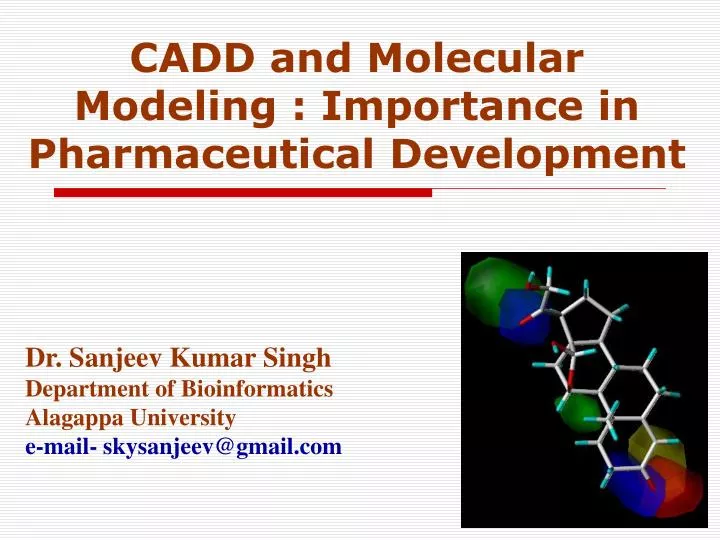
CADD and Molecular Modeling : Importance in Pharmaceutical Development. Dr. Sanjeev Kumar Singh Department of Bioinformatics Alagappa University e-mail- skysanjeev@gmail.com. Working at the Intersection. Structural Biology Biochemistry Medicinal Chemistry Toxicology Pharmacology

E N D
CADD and Molecular Modeling : Importance in Pharmaceutical Development Dr. Sanjeev Kumar Singh Department of Bioinformatics Alagappa University e-mail- skysanjeev@gmail.com
Working at the Intersection • Structural Biology • Biochemistry • Medicinal Chemistry • Toxicology • Pharmacology • Biophysical Chemistry • Information Technology
Structural Biology • Fastest growing area of biology • Protein and nucleic acid structure and function • How proteins control living processes
Medicinal Chemistry • Organic Chemistry • Applied to disease • Example: design new enzyme inhibitor drugs doxorubicin (anti-cancer)
Pharmacology • Biochemistry of Human Disease • Different from Pharmacy: distribution of pharmaceuticals, drug delivery systems
New Ideas From Nature • Natural Products Chemistry • Chemical Ecology • During the next two decades: the major activity in organismal biology • Examples: penicillin, taxol (anti-cancer)
Bio/Chem-informatics • The collection, representation and organisation of chemical data to create chemical information, to which theories can be applied to create chemical knowledge. Aim • To examine how computational techniques can be used to assist in the design of novel bioactive compounds. • To give an idea of how computational techniques can similarly be applied to other emerging areas such as Bio-informatics, Cheminformatics & Pharmainformatics.
Overview • Drug discovery process • How do drugs work? • Overview of Computer-Aided Drug Design
Pharmaceutical/Agrochemical Industry • Identification of novel compounds with useful and commercially valuable biological properties. • vastly complex, • multi-disciplinary task • many stages over extended periods of time • Risk • most novel compounds do not result in a drug. • those that do may cause unexpected, long-term side-effects.
Why CADD…? • Drug Discovery today are facing a serious challenge because of the increased cost and enormous amount of time taken to discover a new drug, and also because of rigorous competition amongst different pharmaceutical companies.
Drug Discovery & Development Identify disease Find a drug effective against disease protein (2-5 years) Isolate protein involved in disease (2-5 years) Scale-up Preclinical testing (1-3 years) Human clinical trials (2-10 years) File IND Formulation File NDA FDA approval (2-3 years)
develop assay 10,000’s compounds lead identification lead optimisation clinical trials 1 drug to market Drug Development Process On average it takes 12 -15 years and costs ~$500 -800 million to bring a drug to market
Technology is impacting this process GENOMICS, PROTEOMICS & BIOPHARM. Potentially producing many more targets and “personalized” targets HIGH THROUGHPUT SCREENING Identify disease Screening up to 100,000 compounds a day for activity against a target protein VIRTUAL SCREENING Using a computer to predict activity Isolate protein COMBINATORIAL CHEMISTRY Rapidly producing vast numbers of compounds Find drug MOLECULAR MODELING Computer graphics & models help improve activity Preclinical testing IN VITRO & IN SILICO ADME MODELS Tissue and computer models begin to replace animal testing
X-ray or Homology Med Chem/Combichem Gene sequence data LibmakerTM Designed libraries Skelgen™ Designed Templates Library synthesis Screening Ligand binding data Pharmacophore Model Automating the CADD Process
Database filtering QSAR Alignment Computer Aided Drug Design (CADD) ADMET Similarity analysis Biophores VHTS diversity selection Combinatorial libraries de novo design Phases of CADD Target discovery Lead discovery Target Identification Target Validation Lead Identification Lead Optimization SAVING 12 – 15 years, Costs: 500 - 800 million US $
How Drugs Work + Substrate Enzyme Enzyme-substrate complex Lock-and-key model
Methodologies and strategies of CADD: • Structure based drug design (SBDD) “DIRECT DESIGN” • Followed when the spatial structure of the target is known. • Ligand based drug design (LBDD) “INDIRECT DESIGN” • Followed when the structure of the target is unknown.
Computer-Aided Drug Design • 3-D target structure unknown (LBDD) • Random screening if no actives are known • Similarity searching • Pharmacophore mapping • QSAR (2D & 3D) etc. • Combinatorial library design etc. • Structure-based drug design (SBDD) • Molecular Docking • De novo design
In Pharmacophore… • Pharmacoporic Studies on ACE inhibitors • Pharmacological Studies on HIV-1RT • Nucleosidic Inhibitors • Non-Nucleosidic Inhibitors • Interaction Energy – Potency Correlation
What is Pharmacophore…? • Pharmacophore model • Set of points in space defining the binding of ligands with target. • Key factors in developing such a model are the determination of functional groups essential for binding, their correspondence from one ligand to another, and the common spatial arrangement of these groups when bound to the receptor The pharmacophore model of HIV protease.
Pharmacophore…..? • “a molecular framework that carries (phoros) the essential features responsible for a drug’s (pharmacon) biological activity” Paul Erlich, early 1990 • “a set of structural features in a molecule that is recognized at a receptor site and is responsible for that molecule’s activity” Peter Gund, 1977
Basic Features • A set of features common to a series of active molecules • What are the features…? • HBD • HBA • +ve &-ve charged groups and • Hydrophobic regions • Functional groups or molecules with similar physical and chemical properties • Bioisosteres - substituents or groups that have chemical or physical similarities and which produce broadly similar biological properties
Pharmacophore model • Set of points in space defining the binding of ligands with target. • Key factors in developing such a model are the determination of functional groups essential for binding, their correspondence from one ligand to another, and the common spatial arrangement of these groups when bound to the receptor.
ACE • Angiotension converting enzyme • Converts angiotensinI to angiotension II • Inhibits bradykinin (vasodilator) • Vasoconstriction
ACE-inhibitor • Orally available & potent drug
ACE distance map • 4 points defined • Five distances defined
Acceptor Charged negative Donor Hydrophobic core
Pharmacophoric Features of Nucleosidic HIV-1RT Inhibitors deoxy nucleoside triphosphate (dNTP) 3'-azido thymidine (AZT) 2',3' dideoxy nucleoside 3'-nitro nucleoside 2',3'- didehydro dideoxy nucleoside MESP contours for nucleosidic drugs. Red coloured contours indicate a value of -.01 for electrostatic potential and yellow contours indicate a value of -0.05
Concluding remarks on Nucleosidic inhibitors • Different substituents at the 3 position show similar sugar ring puckering and only slight differences in nucleosidic base disposition and interactions protein. • MESP plots have clearly indicated that the charge environment of the drugs is complementary to the receptor charge environment.Positive potential areas have been observed in the active site of HIV-1RT where DNA binding occurs. • Pharmacophoric Features of Nucleosidic HIV-1RT Inhibitors. • Arpita Yadav* and Sanjeev Kumar Singh Bioorg. & Med. Chem. 11, 2003, 1801.
Threshold interaction energy of NRTI’s (nucleosidic inhibitors for Reverse transcriptase) to undergo competitive inhibition 3.58 Å 2'3' dideoxy thymidine -13.33 kcal/mol -14.13 kcal/mol AZT -16.71 kcal/mol 3’-Nitro nucleoside -21.30 kcal/mol 2'3'-didehydro 2'3'-dideoxy thymidine -12.39 kcal/mol Correlation of interaction energy with potency
Concluding remarks on interaction energy studies • Correlation graph indicates the requirement of a threshold binding energy ~12 kcal/mol for the drug to be able to undergo competitive inhibition efficiently. Less than this binding energy/ interaction energy will make the drug ineffective or very high concentrations will be required for inhibition of enzyme. Which may lead to cytotoxicity. • vThreshold interaction energy of NRTI’s (nucleosidic inhibitors for Reverse transcriptase) to undergo competitive inhibition • Arpita Yadav* and Sanjeev Kumar Singh Bioorg. & Med. Chem. letts. 14, 2004, 2677-2680
Common binding mode for structurally and chemically diverse non- nucleosidic HIV-1RT inhibitors Pyrrolyl hetro aryl sulfone with lysine
Concluding remarks of Non nucleosidic inhibitors • Conformational study of non-nucleosidic drugs indicated that each drug has a ‘V’- shaped conformation. • Each drug has a -NH group in a position that it can make H- bond with the carbonyl group of lysine 101 in conformity with earlier studies on pyrrolyl hetero aryl sulfone. This indicates the importance of lysine 101 in binding NNRTI’s. • Common binding mode for structurally and chemically diverse non- nucleosidic HIV-1RT inhibitors" Arpita Yadav* and Sanjeev Kumar Singh, THEOCHEM, 723, 2005, 205-209.
DISCO: DIStance COmparisons • Generate some number of low-energy conformations for each active compound • The resulting conformations are represented by the positions of potential pharmacophore points. • Hydrogen-bond donors and acceptors; charged atoms; ring centroids; and centres of hydrophobic regions.
Quantitative Structure-Activity Relationships (QSAR) • A QSAR relates a numerical description of molecular structure or properties to known biological activity • Activity = f (molecular descriptors) • Success of QSAR: right descriptors + right method (form of f ) • A QSAR should be • explanatory (for structures with activity data) • predictive (for structures without activity data) • A QSAR can be used to explain or optimise: • localised properties of molecules such as binding properties • whole molecule properties such as uptake and distribution
3D QSAR • CoMFA and CoMSIA • Molecules are described by the values of molecular fields calculated at points in a 3D grid • The molecular fields are usually steric and electrostatic • Partial least squares (PLS) analysis used to correlate the field values with biological activity • A common pharmacophore is required.
Using the Model • The PLS results are presented as contour plots • Steric Bulk: • Green = Steric Favourable • Yellow = Steric Unfavourable • Electrostatics: • Red = Electronegative Favourable • Blue = Electronegative Unfavourable
3D-QSAR CoMFA Study on Aminothiazole Derivatives as Cyclin Dependent Kinase 2 Inhibitors • In this work we performed CoMFA study carried out on 47 aminothiazole derivatives as inhibitors of this protein kinase. • The models could be usefully employed to design selective CDK2 inhibitors and to find novel scaffolds through screening of chemical databases. Allignment
CoMFA Electrostatic Contours CoMFA Steric Contours • Green contours stand for points where sterically bulkier groups are anticipated to increase the biological activity. • The yellow contours are used to underscore the points where bulkier groups could lower the biological property. • The electrostatic red plots show where the presence of a negative charge is expected to enhance the activity. • The blue contours indicate where introducing or keeping positive charges are expected to better the observed activity. • 3D-QSAR CoMFA Study on Aminothiazole Derivatives as Cyclin Dependent Kinase 2 Inhibitors. Nigus Dessalew, Sanjeev Kumar Singh* and P.V. Bharatam QSAR Comb. Sci., 26(1), 2007, 85-91.
QSAR WORK… • The developed model showed a strong correlative and predictive capability having a cross validated correlation co-efficient of 0.747 for CDK4 and 0.755 for CDK2 inhibitions. • 3D-QSAR CoMFA studies on Indenopyrazole as CDK2 Inhibitors. Sanjeev Kumar Singh*, Nigus Dessalew, and P. V. Bharatam Eur. J. of Med. Chem., 41, 2006, 1310-1319. • The conventional and predictive correlation coefficients were found to be respectively 0.943 and 0.508 for CDK1 and 0.957 and 0.585 for CDK2. • 3D-QSAR CoMFA Study on Oxindole Derivatives as Cyclin Dependent Kinase 1 (CDK1) and Cyclin Dependent Kinase 2 (CDK2) Inhibitors. Sanjeev Kumar Singh*, Nigus Dessalew, and P. V. Bharatam, Med. Chem. 3(1), 2007, 75-84.
Structure Based Drug Design Determine Protein Structure Identify Interaction Sites Discovery or design of molecules that interact with biochemical targets of known 3D structure De Novo Design 3D Database Evaluate Structure Synthesize Candidate Test Candidate Lead Compound
Structure based drug design • Molecular database mining • Compounds with best complementarity to binding site are selected. • DOCK, Autodock, Flex X etc. • De novo drug designing • Virtual modeling and optimization of structure • LUDI, CLIX, CAVEAT, LeapFrog etc.
Structural Targets • 3D structure of target receptors determined by • X-ray crystallography • NMR • Homology modeling • Protein Data Bank • Archive of experimentally determined 3D structures of biological macromolecules
Molecular docking • Virtual screening approach to predict receptor-ligand binding modes • Scoring method used • to detect correct bound conformation during docking process • to estimate binding affinities of candidate molecule after completion of docking
Docking algorithms • Molecular flexibility • both ligand and protein rigid • flexible ligand and rigid protein • both ligand and protein flexible • search algorithm • use to explore optimal positions of the ligand within the active site • scoring function • value should correspond to preferred binding mode • efficiency very important for database searching
Scoring function • Ligand-receptor binding is driven by • Electrostatics (including h-bonding) • Dispersion of vdw’s forces • Hydrophobic interaction • Desolvation of ligand and receptor • Molecular mechanics • Attempt to calculate interaction energy directly
Docking X-ray structure of complex