Download
1 / 1
10 likes | 93 Views
AMYOTROPHIC LATERAL SCLEROSIS: IT’S POSSIBILE TO IDENTIFY A METABOLIC PATTERN ON 18F-FDG-PET? A. Cistaro 1 ; M. Pagani 2; A. Chiò 3; A. Calvo 3; A. Montuschi 3; D. Salmaso 2; F. M. Nobili 4; C. Boffaro 5; G. Carrara5; K. Davari 6; P. Fania 1; C. Casalone 7; G.P. Pescarmona 8; M. C. Valentini 5.

E N D
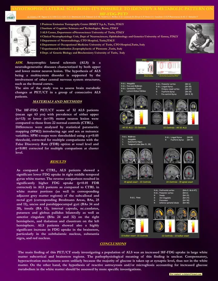
AMYOTROPHIC LATERAL SCLEROSIS: IT’S POSSIBILE TO IDENTIFY A METABOLIC PATTERN ON 18F-FDG-PET? A. Cistaro 1; M. Pagani 2; A. Chiò 3; A. Calvo 3; A. Montuschi 3; D. Salmaso 2; F. M. Nobili 4; C. Boffaro 5; G. Carrara5; K. Davari 6; P. Fania 1; C. Casalone 7; G.P. Pescarmona 8; M. C. Valentini 5 1 Positron Emission Tomography Center IRMET S.p.A., Turin, ITALY 2 Institute of Cognitive Sciences and Technologies, Rome, ITALY 3 ALS Center, Department ofNeuroscience University of Turin, ITALY 4 Clinical Neurophysiology Unit, Dept of Neurosciences, Ophthalmology and Genetics University ofGenoa, ITALY 5 Department of Neuroradiology, CTO Hospital, Turin,ITALY 6 Department of Occupational Medicine University of Turin, CTO Hospital,Turin, Italy 7 Experimental Institution Zooprophylactic of Piemonte ,Turin, Italy 8 Dept. of Genetic Biology and Biochemistry University of Turin, Italy AIM. Amyotrophic lateral sclerosis (ALS) is a neurodegenerative diseases characterized by both upper and lower motor neuron lesion. The hypothesis of ALS being a multisystem disorder is supported by the involvement of other central nervous system structures, such as the frontal cortex. The aim of the study was to assess brain metabolic changes at PET/CT in a group of consecutive ALS patients. MATERIALS AND METHODS The 18F-FDG PET/CT scans of 32 ALS patients (mean age 63 yrs) with prevalence of either upper (n=13) or lower (n=19) motor neuron lesion were compared to those from 22 normal controls (CTRL). Differences were analyzed by statistical parametric mapping (SPM2) introducing age and sex as nuisance variables. SPM t-maps were thresholded using a p<0.05 threshold, corrected for multiple comparisons with the False Discovery Rate (FDR) option at voxel level and p<0.001 corrected for multiple comparison at cluster level. RESULTS As compared to CTRL, ALS patients showed a significant lower FDG uptake in right middletemporal gyrus white matter. The reverse comparison resulted in significantly higher FDG uptake (p<0.01 FDR corrected) in ALS patients as compared to CTRL in white matter portions (as well in corresponding adjacent grey matter regions) of the subcallosal and rectal gyri (corresponding Brodmann Areas, BAs, 25 and 11), uncus and parahippocampal gyri (BAs 34 and 28), insula (BA 13), internal capsula, nc.caudatus, putamen and globus pallidus bilaterally as well as anterior cingulate (BAs 24 and 32) on the right hemisphere, and thalamus and claustrum on the left hemisphere. ALS patients showed also a highly significant increase in FDG uptake in the brainstem, particularly in the subthalamic nucleus, substantia nigra, and red nucleus. CONCLUSIONS The main finding of this PET/CT study investigating a population of ALS was an increased 18F-FDG uptake in large white matter subcortical and brainstem regions. The pathophysiological meaning of this finding is unclear. Compensatory, hyperactivation mechanisms seem unlikely because the majority of glucose is taken up at synaptic level, thus not in the white matter. On the other hand, the hypothesis of reactive astrocytosis and/or microgliosis accounting for increased glucose metabolism in the white matter should be assessed by more specific investigations. For contact: a.cistaro@irmet.com