Download

1 / 25
250 likes | 398 Views
Lecture 1 Overview of early mouse development and methodology nb reading list is at end of notes for this lecture. Vertebrate development – classical models. 1 cm. 100 microns. 1mm. Phylotypic stage. Similar. Similar. Vertebrates are triploblasts.

E N D
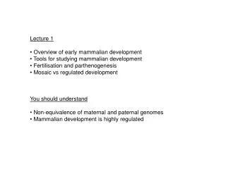
Lecture 1 Overview of early mouse development and methodology nb reading list is at end of notes for this lecture
Vertebrate development – classical models 1 cm 100 microns 1mm Phylotypic stage Similar Similar
Vertebrates are triploblasts Three germ layers, Ectoderm, Mesoderm, and Endoderm (derm=layer), give rise to all cells and tissuesof the developing embryo. Development = origami using layers/sheets of cells
Why study the mouse? • The Victorian mouse fancy movement provided a ready made resource • of inbred strains, variants and mutants • Fast generation time (21 day gestation) • Tissue culture models • Amenable to genetic manipulation
Identity and location Cell Fate Positional information Anterior (Head) Right Dorsal (Back) Ventral (Front) Left Posterior (Tail)
In utero development in mouse occurs over 19-21 days • E (embryo stage) = dpc (days post coitum). Most commonly referred to from 0.5 onwards • as mating takes place at night. • Preimplantation development occurs up to E3.5. All other development occurs • postimplantation.
0 1 2 3 4 Preimplantation Development days Cleavage stages Primitive (primary) endoderm Blastocoel cavity Blastomere Inner cell mass/ Primitive ectoderm Zona pelucida Trophectoderm Activation of embryonic genome
Confusing nomenclature! A ‘derm’ is a cell layer – not a cell type!
Our understanding of the world can only be as good as the state of the art technology we use to measure it – knowledge is relative, not absolute.
Experimental Tools for studying mouse embryos Embryological approaches; • Histological analysis and conventional microscopy • In vitro culture of preimplantation stages and in some cases postimplantation stages. • Cell fate mapping (dyes and now tagged loci)
Embryological approaches; • Embryo manipulation/transplantation • Chimera formation and embryo aggregation. e.g. tetraploid chimeras for testing gene function in extraembryonic vs embryonic lineages. • Cell culture models Embryonic stem (ES) cells
Molecular embryology; • Gene expression profiling of embryos, dissected fragments, derivative tissue culture cell lines and single cells. • In situ hybridization Sections Wholemount • Immunohistochemistry Eed +Nanog Oct4 +Eed
Genetic approaches; • Classical mouse mutants Brachyury mouse with short tail is dominant mutation in gene for transcription factor required for mesoderm formation. • Genetic screens Chemical (ENU) mutagenesis – requires lengthy genetic mapping and cloning to identify mutated locus Insertional or ‘gene trap’ mutagenesis in ES cells – can go directly to gene of interest Antibiotic resistance marker Reporter gene PolyA signal IRES SA Wild-type and Nodal (d/d) mutant embryos with staining for markers of primitive streak (brown) and ectoderm (dark blue). SD
Genetic manipulation in mouse; • Production of transgenic mice • by pronuclear injection of DNA • Production of genetically modified mice • by transferring ES cells to recipient embryo - Gene manipulation using homologous recombination in ES cells - Inject modified cells into Recipient embryo to produce chimeric animal that transmits donor genome through the germ-line. - Gene construct injected into male pronucleus of 1-cell embryo - DNA integrates randomly at single site, usually multicopy
Genetic manipulation in mouse; • Gene targeting in embryonic stem (ES) cells
Negative selectable Marker gene Positive selectable Marker gene X X Genetic manipulation in mouse; Conventional gene knockout strategy (replacement vector) Knock-out (or Knock-in)
X X + site specific recombinase (Cre or Flp) + Genetic manipulation in mouse; Conditional gene knockout strategy; Negative selectable Marker gene Recombinase recognition sequence Positive selectable Marker gene
Genetic manipulation in mouse; Conditional gene knockout strategy; Transgenic mouse expressing site specific recombinase in tissue specific pattern Homozygous conditional allele X Analyse phenotype in F1 embryos or adults Examples of recombinase driver transgenics; - Cre recombinase driven by Nanog promoter - Estrogen receptor-Cre recombinase fusion driven by constitutive promoter. Addition of Tamoxifen to drinking water triggers nuclear translocation of recombinase giving temporal control of gene deletion.
Reading list Textbook; Principles of Development, Lewis Wolpert and Cheryl Tickle. Review papers; Lecture 1 -3 Alexandre (2001) International Journal of Developmental Biology 45, p457-467 Rossant (2001) Stem Cells 19, p477-82 Yamanaka et al, (2006). Developmental Dynamics 235, p2301-2314 Katsuyoshi and Hamada, (2012) Development 139, p3-14 Lecture 4 and 5 Arnold and Robertson (2009) Nature reviews Molecular cellular biology, 10, p91-103 Robb and Tam (2004) Seminars in Cell and Developmental biology 15, p43-54 Hayashi et al (2007) Science 316, p394-396. Hashimoto and Hamada (2010) , CurrOpin Genet Dev 20, p433-7 Hanna et al (2010) Cell 143, p508-525. Yamanaka and Blau (2010) Nature 465, p704-712
New innovations in ES cell manipulation (optional if time permits)
Genetic manipulation in mouse; ZFN, TALEN and CrispR/cas systems; • TALE effector proteins secreted by Xanthomonas bacteria in order to activate host plant gene expression that aids infection. • Modular composition of sequence specific binding domains comprising 33-34 amino acids with positions 12 and 13 being highly variable. • Can be used to construct designer Transcription Activator Like Effector Nuclease (TALEN) to introduce DNA breaks at defined target sequence. • Provides substrate for error prone repair or HR using recombinant DNA template for custom modification. • Cys2-His2 zinc finger domain contacts 3bp of sequence in major groove with varying levels of selectivity. • Can use as modular component to get sequence specific targeting of Fokl restriction endonuclease monomer. Cleavage requires targeting second monomer to other strand to generate functional Fokl dimer. • Provides substrate for error prone repair or HR using recombinant DNA template for custom modification.
Genetic manipulation in mouse; ZFN, TALEN and CrispR/cas systems; (Trans-encoded CRISPR RNA) PAM site • RNA mediated bacterial defense against viral or plasmid DNA. • Type II system adapted for genome engineering in many organisms. • Can use cas9 intrinsic nuclease to introduce ds break or ss nick. • Provides substrate for error prone repair orHR using recombinant DNA template for custom modification. • Can also mutate directly by injection into zygote. • Partially circumvents requirement for highly recombinogenic cell such as ES cell.