Download
1 / 53
540 likes | 669 Views
Ch. 31 – Plant Structure, Growth and Differentiation. Plant Body. Root system Underground Anchor and absorb Shoot system Vertical stem, leaves (flowers, fruits w/seeds) photosynthesis. Fig. 35-2. Reproductive shoot (flower). Apical bud. Node. Internode. Apical bud. Shoot system.

E N D
Plant Body • Root system • Underground • Anchor and absorb • Shoot system • Vertical stem, leaves (flowers, fruits w/seeds) • photosynthesis
Fig. 35-2 Reproductive shoot (flower) Apical bud Node Internode Apical bud Shoot system Vegetative shoot Blade Leaf Petiole Axillary bud Stem Taproot Lateral branch roots Root system
Plant Cells and Tissues • Ground tissue system – majority • Photosynthesis, storage, support • Vascular tissue system • Conduction, strength, support • Dermal tissue system • Covering, protection All 3 are Interconnected throughout the plant
Fig. 35-8 Dermal tissue Ground tissue Vascular tissue
Ground Tissue System • Parenchyma, collenchyma, sclerenchyma tissue • Primary cell wall – secreted by growing cell; stretches and expands as cell grows • Secondary cell wall – secreted when cell stops growing; thick and strong (inside primary)
Parenchyma • Living, metabolizing • Most common • Soft parts • Function • Photosynthesis – green chloroplasts • Storage – starch, oil, water, salt • Secretion – resins, tannins, hormones, enzymes, nectar • Can differentiate if plant injured (i.e. xylem cells)
Fig. 35-10a Parenchyma cells in Elodea leaf, with chloroplasts (LM) 60 µm
Collenchyma • Flexible, structural support (nonwoody parts) • Elongated cells • Alive at maturity • Primary CW – unevenly thick, thicker in corners • Near stem surface, leaf veins
Fig. 35-10b 5 µm Collenchyma cells (in Helianthus stem) (LM)
Sclerenchyma • Structural support • Primary and secondary CW (strong and hard, extreme thickening, so can’t stretch, elongate) • Cells dead at maturity • 2 types: • Sclereids – variable shape, nut shells, pits of stone fruits, pears gritty (clusters of sclereids) • Fibers – long, tapered – patches, clumps; wood, inner bark, leaf veins
Fig. 35-10c 5 µm Sclereid cells in pear (LM) 25 µm Cell wall Fiber cells (cross section from ash tree) (LM)
Vascular tissue • Embedded in ground tissue • Transport • Xylem and phloem
Xylem • Conducts water, dissolved nutrient minerals roots stems, leaves • Support • Angiosperms – • tracheids, vessel elements - conduct • parenchyma cells - storage • fibers - support
Tracheids and vessel elements • Dead at maturity hollow, CW remain • Tracheids – long, tapering, patches/clumps; water passes from 1 tracheid to another by pits (thin areas where sec. wall did not form) • Vessel elements – larger in diameter than tracheid; end walls have perforations; stacked water goes between; stack = vessel; pits in side walls for lateral water transport
Fig. 35-10d 100 µm Vessel Tracheids Pits Tracheids and vessels (colorized SEM) Perforation plate Vessel element Vessel elements, with perforated end walls Tracheids
Phloem • Conducts food • Support • Angiosperms • Sieve tube members, companion cells – conduct • Fibers – support • Parenchyma cells
Sieve tube members • Conduct food in solution • Joined end-to-end long tubes • CW ends = sieve plates; cytoplasm extends between cells • Living at maturity – many organelles shrink/disintegrate • Can function w/o nuclei
Companion cells • Adjacent to each sieve tube member (stm) • Assists stm • Living w/ nucleus – directs activities of both cells • Plasmodesmata between stm and companion • Helps move sugar into stm
Fig. 35-10e Sieve-tube elements: longitudinal view (LM) 3 µm Sieve plate Sieve-tube element (left) and companion cell: cross section (TEM) Companion cells Sieve-tube elements Plasmodesma Sieve plate 30 µm 10 µm Nucleus of companion cells Sieve-tube elements: longitudinal view Sieve plate with pores (SEM)
Dermal tissue system • Epidermis and periderm • Protective covering • Herbaceous – single layer = epidermis • Woody – epidermis splits w/ growth • Periderm – layers thick, under epidermis; replaces epidermis in stems, roots, composing outer bark
Epidermis • Unspecialized dermal cells • Special guard cells + trichomes • Single layer, flat cells • Usually no chloroplasts transparent • Allow light through
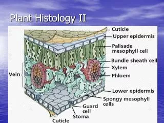
Fig. 35-18a Key to labels Dermal Ground Cuticle Sclerenchyma fibers Vascular Stoma Upper epidermis Palisade mesophyll Spongy mesophyll Bundle- sheath cell Lower epidermis Cuticle Xylem Vein Phloem Guard cells (a) Cutaway drawing of leaf tissues
Fig. 35-18b Guard cells Stomata pore 50 µm Epidermal cell (b) Surface view of a spiderwort (Tradescantia) leaf (LM)
Fig. 35-18c Upper epidermis Key to labels Palisade mesophyll Dermal Ground Vascular Spongy mesophyll Lower epidermis 100 µm Air spaces Guard cells Vein (c) Cross section of a lilac (Syringa) leaf (LM)
Cuticle • Aerial parts • Secreted by epidermal cells • Waxy – water loss • Slows diffusion of CO2 – stomata help • Stomata • Open – day – photosynthesis, evaporative cooling • Closed – night • Closed in day if drought
Trichomes • Outgrowths or hairs • Many shape, sizes, functions • Ex: • Roots hairs – increase SA • Salty env. – remove excess salt • Aerial parts – increase light reflection, cooler • Protections – stinging nettles
Growth at Meristems • Cell division • Increase # cells • Cell elongation • Vacuole fills, increase pressure on CW, expands • Cell differentiation • Specialize into cell types • Meristems = where plant cells divide, mitosis • No differentiation
2 kinds of Growth • Primary growth • Increase stem, root length • All plants, soft tissues • Secondary growth • Increase width • Gymnosperms, woody dicots • Wood + bark
Fig. 35-11 Primary growth in stems Epidermis Cortex Shoot tip (shoot apical meristem and young leaves) Primary phloem Primary xylem Pith Lateral meristems: Vascular cambium Secondary growth in stems Cork cambium Periderm Axillary bud meristem Cork cambium Cortex Primary phloem Pith Primary xylem Secondary phloem Root apical meristems Secondary xylem Vascular cambium
Primary growth • Increase in length • Apical meristem – tips of roots + shoots (buds) • Buds = dormant embryonic shoot (develop into branches next spring • Root tip • Root cap – protective layer of cells, covers root tip • Root apical meristem – directly behind root cap • Cell elongation – behind meristem, push tip ahead, some differentiation
Fig. 35-13 Cortex Vascular cylinder Epidermis Key to labels Zone of differentiation Root hair Dermal Ground Vascular Zone of elongation Apical meristem Zone of cell division Root cap 100 µm
Fig. 35-14a1 Epidermis Key to labels Cortex Dermal Ground Endodermis Vascular Vascular cylinder Pericycle Xylem 100 µm Phloem (a) Root with xylem and phloem in the center (typical of eudicots)
Fig. 35-14a2 (a) Root with xylem and phloem in the center (typical of eudicots) Endodermis Key to labels Pericycle Dermal Ground Vascular Xylem Phloem 50 µm
Fig. 35-14b Epidermis Cortex Endodermis Vascular cylinder Key to labels Pericycle Dermal Core of parenchyma cells Ground Vascular Xylem Phloem 100 µm (b) Root with parenchyma in the center (typical of monocots)
Shoot apex = terminal bud • Shoot meristem • Give rise to leaf primordia and bud primordia
Fig. 35-16 Leaf primordia Shoot apical meristem Young leaf Developing vascular strand Axillary bud meristems 0.25 mm
Fig. 35-17a Phloem Xylem Sclerenchyma (fiber cells) Ground tissue connecting pith to cortex Pith Key to labels Cortex Epidermis Dermal Vascular bundle Ground Vascular 1 mm (a) Cross section of stem with vascular bundles forming a ring (typical of eudicots)
Fig. 35-17b Ground tissue Epidermis Key to labels Vascular bundles Dermal Ground Vascular 1 mm (b) Cross section of stem with scattered vascular bundles (typical of monocots)
Secondary Growth • Increase in width • Make secondary tissues: sec. xylem, sec. phloem, periderm • Lateral meristem – cells divide, not elongate • 2 types: • Vascular cambium • Between wood and bark • Make sec. xylem (wood) + sec. phloem (inner bark)
Fig. 35-20 Growth Vascular cambium Vascular cambium X X C P P Secondary phloem Secondary xylem X C P X C X P C C C C X C C C After one year of growth After two years of growth C C C
Fig. 35-22 Growth ring Vascular ray Heartwood Secondary xylem Sapwood Vascular cambium Secondary phloem Bark Layers of periderm
Cork cambium • In outer bark • Form cork to outside +parenchyma (storage) • Periderm = cork, parenchyma, cork cambium
Bark – outermost covering of woody stems • Everything outside of vascular cambium • 2 regions: • Living inner bark of secondary phloem • Mostly dead outer bark of periderm