Download
1 / 46
460 likes | 470 Views
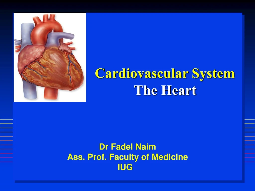
Cardiovascular System The Heart. Dr Fadel Naim Ass. Prof. Faculty of Medicine IUG. Size of Heart. Average Size of Heart 14 cm long 9 cm wide. Location of Heart. Inside thoracic cavity posterior to sternum medial to lungs anterior to vertebral column

E N D
Cardiovascular SystemThe Heart Dr Fadel Naim Ass. Prof. Faculty of Medicine IUG
Size of Heart • Average Size of Heart • 14 cm long • 9 cm wide
Location of Heart • Inside thoracic cavity • posterior to sternum • medial to lungs • anterior to vertebral column • apex tipped toward the left; base superior • base lies beneath 2nd rib • apex at 5th intercostal space • lies upon diaphragm
Functions of the Heart • Center of the cardiovascular system, the heart. • Connects to blood vessels that transport blood between the heart and other body tissues. • arteries carry blood away from the heart • veins carry blood back to the heart • Arteries carry blood high in oxygen. • (except for the pulmonary arteries) • Veins carry blood low in oxygen. • (except for the pulmonary veins) • Arteries and veins entering and leaving the heart are called the great vessels.
Characteristics and Functions of the Heart • Ensures the unidirectional flow of blood through both the heart and the blood vessels. • Backflow of blood is prevented by valves within the heart. • Acts like two independent, side-by-side pumps that work independently but at the same rate. (double circuit) • one directs blood to the lungs for gas exchange • the other directs blood to body tissues for nutrient delivery
Characteristics and Functions of the Heart • Develops blood pressure through alternate cycles of heart wall contraction and relaxation. • Minimum blood pressure is essential to push blood through blood vessels to the body tissues for nutrient and waste exchange.
artery vein capillaries Pulmonary & Systemic Circuits
Pulmonary and Systemic Circuits • The pulmonary circuit consists of the chambers on the right side of the heart (right atrium and ventricle) as well as the pulmonary arteries and veins. • conveys blood to the lungs via pulmonary arteries • to reduce carbon dioxide and replenish oxygen levels in the blood • Blood returns to the heart in pulmonary veins
Pulmonary and Systemic Circuits • Blood returns to the left side of the heart, where it then enters the systemic circuit. • The systemic circuit consists of the chambers on the left side of the heart (left atrium and ventricle), along with all the other named blood vessels. • carries blood to all the peripheral organs and tissuesof the body
Pulmonary and Systemic Circuits • Oxygenated blood from the left side of the heart is pumped into the aorta • the largest systemic artery in the body • then into smaller systemic arteries. • Gas exchange in tissues occurs from capillaries. • Systemic veins then carry deoxygenated blood (high in carbon dioxide) and waste products. • Most veins merge and drain into the superior and inferior venae cavae • drain blood into the right atrium. • There, the blood enters the pulmonary circuit, and the cycle repeats .
Pericardium • Fibrous, serous sac • Contains the heart • In the mediastinum • Held in place by connective tissue • The external wall of the great vessels’ superior to the heart • diaphragm inferior. • Restricts heart movements • Prevents the heart fromoverfilling with blood.
Pericardium • Outer portion • tough, dense connective tissue • called the fibrous pericardium. • attached to both the sternum and the diaphragm • Inner portion • thin, double-layered serous membrane • called the serous pericardium. • parietal layer • visceral layer
Heart Wall Structure • Three distinctive layers: • external epicardium • middle myocardium • internal endocardium • Epicardium • outermost heart layer • also known as the visceral layer of serous pericardium. • Simple squamous epithelium underlined by fat • As we age, more fat is deposited in the epicardium • this layer becomes thicker and more fatty.
Heart Wall Structure • Myocardium • middle layer of the heart wall • composed chiefly of cardiac muscle tissue. • thickest of the three heart wall layers. • lies deep to the epicardium and superficial to the endocardium • Endocardium • covers internal surface of the heart and the external surfaces of the heart valves • thin endothelium • areolar CT under the endothelium
Functions of the Fibrous Skeleton of the Heart • Located between the atria and the ventricles • Formed from dense irregular connective tissue. • separates the atria and ventricles • anchors heart valves by forming supportive rings at their attachment points • provides electrical insulation between atria and ventricles • ensures that muscle impulses are not spread randomly throughout the heart • prevents all of the heart chambers from beating at the same time • Provides a rigid framework for the attachment of cardiac muscle tissue.
External Anatomy of the Heart • Chambers: • four hollow chambers: • two smaller atria • two larger ventricles. • Atria • thin-walled, located superiorly. • anterior part of each atrium is a wrinkled, flaplike extension called an auricle • Atria receive blood through both circulatory circuits. • right atrium receives blood from the systemic circuit • left atrium receives blood from the pulmonary circuit
External Anatomy of the Heart • Blood that enters an atrium is passed to the ventricle on the same side of the heart. • Ventricles • the inferior chambers. • Two large arteries, the pulmonary trunk and the aorta exit the heart at the basal surface. • The pulmonary trunk carries blood from the right ventricle into the pulmonary circuit. • The aorta conducts blood from the left ventricle into the systemic circuit
External Anatomy of the Heart • Atria are separated from the ventricles externally by coronary sulcus (or atrioventricular sulcus) • extends around the circumference of the heart. • On both the anterior and posterior surfaces of the heart, the anterior interventricular sulcus and the posterior interventricular sulcus are located between the left and right ventricles. • These sulci extend inferiorly from the coronary sulcus toward the heart apex.
Internal Anatomy of the Heart • There are four heart chambers: • right atrium • right ventricle • left atrium • left ventricle • Each plays a role in the continuous process of blood circulation. • Valves permit the passage of blood in one direction and prevent its backflow.
Left vs. Right Ventricle Left: high pressure pump - Right: low pressure pump right chamber is thinner walled than left Ventricles separated by interventricular septum
Structure and Function of Valves = Mitral valve 4 sets of valves Prevent backflow of blood Close passively under blood pressure Heart sounds produced by valve closure
Support for AV valves: valves are restrained by chordae tendinae which are in turn attached to papillary muscles (prevention of backflow!) picture taken from R ventricle, looking toward R atrium
Mitral Valve Prolapse • Most common cardiac variation (5-10% of population) • Mitral valve cusps do not close properly • Regurgitation during left ventricular systole Not life threatening; may be lifestyle threatening
Blood flow pattern through the heart • Blood enters right atrium • Passes tricuspid valve into right ventricle • Leaves by passing pulmonary semilunar valves into pulmonary trunk and to the lungs to be oxygenated • Returns from the lung by way of pulmonary veins into the left atrium • From left atrium past bicuspid valve into left ventricle • Leaves left ventricle past aortic semilunar valves into aorta • Distributed to rest of the body
Coronary Circulation • Left and right coronary arteries travel in the coronary sulcus (atrioventricular groove) of the heart to supply the heart wall. • the only branches of the ascending aorta • Located immediately superior to the aortic semilunar valve. • The right coronary artery typically branches into the • marginal artery • supplies the right border of the heart • posterior interventricular artery • supplies both the left and right ventricles
Coronary Circulation • Left coronary artery typically branches into the anterior interventricular artery. • also called the left anterior descending artery • supplies the anterior surface of both ventricles and most of the interventricular septum • Circumflex artery. • supplies the left atrium and ventricle • Arterial pattern can vary greatly among individuals.
Risk Factors for CAD • High blood cholesterol • High blood pressure • Smoking • Obesity • Diabetes mellitus • Type “A” personality • Sedentary lifestyle
Myocardial Infarction (MI) • ~ 1.3 Mio MIs / year in US • Most commonly due to severe CAD (coronary thrombosis) • Ischemic tissue degenerates → nonfunctional area = infarct