Download
1 / 15
160 likes | 404 Views
Biology 232 – Physiology & Anatomy 1 Additional Slides for Lecture Exam #3 (With guided notes… be sure to look in the notes sections for PowerPoint in this document… these will be very helpful!). Wilder Penfield 1891 – 1976

E N D
Biology 232 – Physiology & Anatomy 1 Additional Slides for Lecture Exam #3 (With guided notes… be sure to look in the notes sections for PowerPoint in this document… these will be very helpful!)
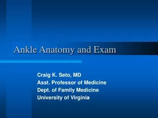
Wilder Penfield 1891 – 1976 Physician and Neuroscientist who mapped the brain in what became the “homunculus”
Figure 12.9: Motor and sensory areas of the cerebral cortex, p. 438. Motor Sensory Shoulder Trunk Trunk Knee Neck Head Leg Hip Hip Arm Arm Elbow Elbow Forearm Wrist Hand Hand Fingers Fingers Thumb Thumb Eye Neck Nose Brow Face Eye Lips Genitals Toes Face Teeth Gums Lips Jaw Tongue Jaw Tongue Pharynx Swallowing Motor cortex (precentral gyrus) Intra- abdominal
Figure 12.5: Ventricles of the brain, p. 434. Lateral ventricle Lateral ventricle Third ventricle Anterior horn Septum pellucidum Posterior horn Third ventricle Cerebral aqueduct Inter- ventricular foramen Cerebral aqueduct Fourth ventricle Inferior horn Median aperture Fourth ventricle Lateral aperture Central canal Central canal (a) Anterior view (b) Left lateral view
Figure 12.26: Formation, location, and circulation of CSF, p. 466. Superior sagittal sinus Superior cerebral vein Arachnoid villus Choroid plexus Cerebrum covered with pia mater Subarachnoid space Arachnoid mater Meningeal dura mater Septum pellucidum Periosteal dura mater Great cerebral vein Corpus callosum Tentorium cerebelli Interventricular foramen Straight sinus Confluence of sinuses Third ventricle Cerebellum Pituitary gland Choroid plexus Cerebral aqueduct Cerebral vessels that supply choroid plexus Lateral aperture Fourth ventricle Median aperture Central canal of spinal cord Spinal dura mater Inferior end of spinal cord Filum terminale (inferior end of pia mater) (b)
Figure 12.31b: Anatomy of the spinal cord, p. 473. Posterior median sulcus Gray commissure Posterior funiculus Dorsal (posterior) horn Gray matter Ventral (anterior) horn White columns Anterior funiculus Lateral horn Lateral funiculus Dorsal root ganglion Spinal nerve Central canal Dorsal root Anterior median fissure Ventral root Pia mater Arachnoid Spinal mater (b)
Figure 13.12a: Dermatomes, p. 518. C2 C3 C4 C5 T1 T2 T3 T4 T2 T2 T5 T6 T7 T8 C5 C5 T9 T10 C6 C6 T11 T12 L1 L1 C6 C6 S2 C7 C7 C8 S3 C8 L2 L2 L3 L3 L4 L4 L5 L5 S1 S1 (a)
Figure 13.12b: Dermatomes, p. 518. C2 C3 C4 C5 C6 C7 C8 T1 C5 T2 T3 T4 T5 T6 T7 T8 T9 T10 C6 C6 T11 T12 C7 C7 L1 S1 L2 C8 C8 L3 S2 L5 L4 S3 S4 S5 S1 S2 S2 S1 L1 L2 L5 L5 L3 L4 L4 L4 L5 L5 S1 (b)
Figure 14.2: Comparison of somatic and autonomic nervous systems, p. 534. Central nervous system Peripheral nervous system Effector organs Acetylcholine Somatic nervous system Skeletal muscle Acetylcholine Norepinephrine Smooth muscle (e.g., in gut) Ganglion Sympathetic division Acetylcholine Epinephrine and norepinephrine Autonomic nervous system Blood vessel Glands Adrenal medulla Acetylcholine Cardiac muscle Para- sympathetic division Ganglion Key: = Preganglionic axons (sympathetic) = Postganglionic axons (sympathetic) = Myelination = Preganglionic axons (parasympathetic) = Postganglionic axons (parasympathetic)
Figure 14.3: Overview of the subdivisions of the ANS, p. 536. Parasympathetic Sympathetic Eye Eye Brain stem Salivary glands Skin* Cranial Salivary glands Sympathetic ganglia Heart Cervical Lungs Lungs T1 Heart Stomach Thoracic Pancreas Stomach Liver and gall- bladder Pancreas L1 Adrenal gland Liver and gall- bladder Lumbar Bladder Bladder Genitals Genitals Sacral
Marshall Hall 1790 – 1857 English physiologist who first advanced the theory of how the reflex arc worked.
Figure 13.14: The basic components of all human reflex arcs, p. 521. Spinal cord (in cross section) Stimulus Sensory neuron 2 Integration center 3 Receptor 1 Interneuron Motor neuron 4 Skin Effector 5
Figure 14.1: Place of the ANS in the structural organization of the nervous system, p. 533. CNS PNS Motor division Sensory division Sympathetic division Autonomic nervous system Somatic nervous system Parasympathetic division
Figure 14.8: Referred pain, p. 543. Heart Lungs and diaphragm Liver Gallbladder Gallbladder Heart Liver Appendix Stomach Pancreas Small intestine Ovaries Colon Kidneys Urinary bladder Ureters