Download
1 / 59
660 likes | 1.03k Views
Optimization of Gamma Knife Radiosurgery. Michael Ferris, Jin-Ho Lim University of Wisconsin, Computer Sciences David Shepard University of Maryland School of Medicine Supported by Microsoft, NSF and AFOSR. Overview. Details of machine and problem Optimization formulation modeling dose

E N D
Optimization of Gamma Knife Radiosurgery Michael Ferris, Jin-Ho Lim University of Wisconsin, Computer Sciences David Shepard University of Maryland School of Medicine Supported by Microsoft, NSF and AFOSR
Overview • Details of machine and problem • Optimization formulation • modeling dose • shot/target optimization • Results • Two-dimensional data • Real patient (three-dimensional) data
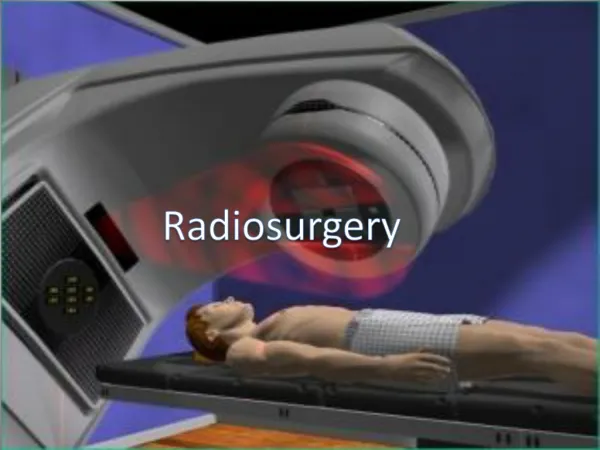
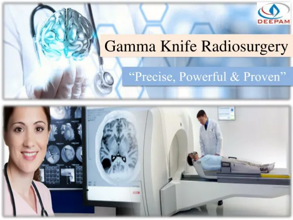
201 cobalt gamma ray beam sources are arrayed in a hemisphere and aimed through a collimator to a common focal point. The patient’s head is positioned within the Gamma Knife so that the tumor is in the focal point of the gamma rays.
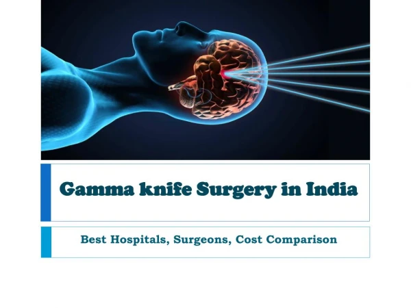
What disorders can the Gamma Knife treat? • Malignant brain tumors • Benign tumors within the head • Malignant tumors from elsewhere in the body • Vascular malformations • Functional disorders of the brain • Parkinson’s disease
Gamma Knife Statistics • 120 Gamma Knife units worldwide • Over 20,000 patients treated annually • Accuracy of surgery without the cuts • Same-day treatment • Expensive instrument
How is Gamma Knife Surgery performed? Step 1: A stereotactic head frame is attached to the head with local anesthesia.
Step 2: The head is imaged using a MRI or CT scanner while the patient wears the stereotactic frame.
Step 3: A treatment plan is developed using the images. Key point: very accurate delivery possible.
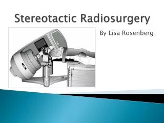
Step 4: The patient lies on the treatment table of the Gamma Knife while the frame is affixed to the appropriate collimator.
Step 5: The door to the treatment unit opens. The patient is advanced into the shielded treatment vault. The area where all of the beams intersect is treated with a high dose of radiation.
Treatment Planning • Through an iterative approach we determine: • the number of shots • the shot sizes • the shot locations • the shot weights • The quality of the plan is dependent upon the patience and experience of the user
Inverse Treatment Planning • Develop a fully automated approach to Gamma Knife treatment planning. • A clinically useful technique will meet three criteria: robust, flexible, fast • Benefits of computer generated plans • uniformity, quality, faster determination
Computational Model • Target volume (from MRI or CT) • Maximum number of shots to use • Which size shots to use • Where to place shots • How long to deliver shot for • Conform to Target (50% isodose curve) • Real-time optimization
Dose calculation • Measure dose at distance from shot center in 3 different axes • Fit a nonlinear curve to these measurements (nonlinear least squares) • Functional form from literature, 10 parameters to fit via least-squares
MIP Approach Choose a subset of locations from S
Features of MIP • Large amounts of data/integer variables • Possible shot locations on 1mm grid too restrictive • Time consuming, even with restrictions and CPLEX • but ... have guaranteed bounds on solution quality
Iterative approach • Approximate via “arctan” • First, solve with coarse approximation, then refine and reoptimize
Difficulties • Nonconvex optimization • speed • robustness • starting point • Too many voxels outside target • Too many voxels in the target (size) • What does the neurosurgeon really want?
Iterative Approach • Rotate data (prone/supine) • Skeletonization starting point procedure • Conformity subproblem (P) • Coarse grid shot optimization • Refine grid (add violated locations) • Refine smoothing parameter • Round and fix locations, solve MIP for exposure times
Status • Automated plans have been generated retrospectively for over 30 patients • The automated planning system is now being tested/used head to head against the neurosurgeon • Optimization performs well for targets over a wide range of sizes and shapes
Environment • All data fitting and optimization models formulated in GAMS • Ease of formulation / update • Different types of model • Nonlinear programs solved with CONOPT (generalized reduced gradient) • LP’s and MIP’s solved with CPLEX
tumor brain
Patient 2 - Axial slice 15 shot manual 12 shot optimized