Download
1 / 26
300 likes | 321 Views
Electrical Injury. Electrical Injury. In the U.S. 52,000 admissions/yr 3-8 % of all burn unit admissions May-Sept lightning related. Decrease in incidence due to GFCIs. Electrical Injury - Epidemiology. Ages 15-44 yrs. High voltage mostly occupational injury 20% Children

E N D
Electrical Injury • In the U.S. 52,000 admissions/yr • 3-8 % of all burn unit admissions • May-Sept lightning related. • Decrease in incidence due to GFCIs
Electrical Injury - Epidemiology • Ages 15-44 yrs. • High voltage mostly occupational injury • 20% Children • Low voltage injuries in toddlersM:F 1.7:1 • High voltage injuries in adolescents97% male
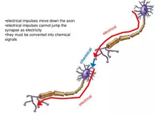
Electrical Injury - Pathophysiology • Electrical – tetany, arrhythmia • Thermal – burns, coagulation • Mechanical – fractures, dislocation
Ohm’s Law I= V/R I= current V= voltage R= resistence
Joule’s Law E=I²RT E= energy I= current R= Resistence T= time
Electrical Injury - Pathophysiology Current pathwaydefines resistence - Vertical higher incidence of complication - Hand – to – hand pathway - Below symphysis, stradle pathway
Electrical Injury - Classification • High (>1000 Volt) vs. low (<1000 Volt) voltage • Direct (lightning) vs. alternating (50 Hz) current • Arc injury (high temperature), flashover
Cardiovascular Involvment • Mostly in vertical injury • DC – Asystole • AC • High VF/ VT, asystole • Low ectopic beats, AF, tachycardia, bradycardia, ECG changes • Coagulation necrosis, coronary spasm, MI
Respiratory Involvement • Tetany of respiratory muscle • Brain stem injury • May induce hypoxia, acidosis cardiac arrest
Nervous System • Immediate - loss of consciousness, amnesia • Early - intracranial hemorrhage, vertebral fractures • Late - ALS, transverse myelitis, ascending paralysis • Peripheral neuropathy, RSD
Vascular Injury • Large arteries – medial necrosis, aneurisms • Small vessels – intimal injury, coagulation necrosis • Secondary to compartment syndrome
Limb Injury • Dislocations and fractures • Coagulation of blood vessels • Muscle ischemia and edema • Compartment syndrome • Thermal injury from bone heating • Infection clostridial, streptococcal
Other Injuries • GI – ileus, stress ulcers, direct injury • Ophthalmic – cataract, iridiocyclitis, autonomic injury • Otologic – tympanic membrane perforation, vertigo, sensoryneural injury
Low Voltage 77% 0-5 YO 60% extremity 40% oral commisure No mortality Complete functional recovery High Voltage 76% 11-18 YO 33% limb amputations 30% deep muscles 12% fasciotomy/ escharotomy No mortality Injury Characteristics
Electrical Injury - Management • Combined ATLS + ACLS protocols • Cardiac monitoring for 24 hrs if LOC, ECG changes or arrhythmias • IM dT • IV H2 - blockers
Electrical Injury – Resuscitation • 1.7 X Parkland formula or 9 ml/kg/%TBSA • Urine output 70 - 100 ml/hour • Clearance of any pigment in urine • Bicarbonate - blood pH > 7.45 • Osmotic diuresis – IV MANNITOL 25 gr
Electrical Injury – Wound Managemant “True” high tension • Sharply demarcated • Always full thickness • Leathery appearence
Electrical injury – Wound Management • Primary resuscitation. • Early exploration and debridment • “Second look” in 24-48 hrs –definitive Tx • Primary closure • Coverage • Amputation • “Progressive necrosis” theory
Wound Management – Extremities • Frequent envolvement of the hand • Exit point in one or both legs • Arc injury in distal fore arm or axilla
Wound Management – Extremities • Initial assessment usually predicts outcome: • Depth of burns • Ischemia • Anasthesia • Flexion position • Muscle viability- response to electrocautery
Wound Management – Extremities • Exploration - large volume underlying necrotic area • Full thickness burns • Proximal periosseous myonecrosis • Retained questionable tissue may lead to contamination and further compromise
Wound Management - Scalp • Saucer shaped, deapest in the middle • Delayed Tx osteomyelitis and epidural abscess • Debridment of soft tissue, outer cortical bone and skin grafting • Full thicknss skull - devitalization & flap coverage
Wound Management – Trunk & perineum • Suspect visceral injury • Lung – Atelectasis and edema • Abdomen – consider as penetrating wound • Perineum –urinary and bowel diversion & debridment +STSG
Electrical Injury -Summery • סוג הפגיעה ומיקומה • טיפול ראשוני לפי פרוטוקולים ACLSו- ATLS • החייאת נוזלים • אקספלורציה והטרייה מוקדמים • טיפול דפיניטיבי מוקדם – בכל שיטות השחזור המקובלות