Download
1 / 9
100 likes | 189 Views
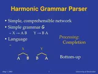
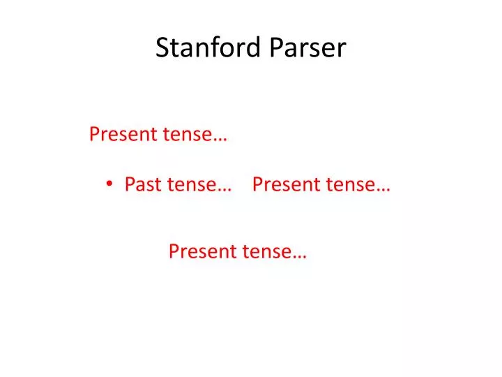
Stanford Parser. Past tense…. Present tense…. Present tense…. Present tense…. Confusion Matrix. Previous findings.

E N D
Stanford Parser • Past tense… Present tense… Present tense… Present tense…
Previous findings • In the left upper lobe, a triangular nodule is present on series 5 image 5. It also was present on the study from 10/17/02. It appears slightly more triangular in shape on the current study but has not changed significantly between exams.
Previous findings • There appears to be a small endoluminal mass in the superior esophagus just caudal to the oropharynx. This was present on the previous exam and is stable.
Previous findings • It currently measures 42 x 37 mm, decreased from 49 mm on the previous study.
Previous findings • The confluent lymph node mass in the low right paratracheal space extending across midline that measured 37 x 19 mm on the previous exam is 35 x 20 mm on the current study. Itappears to have less craniocaudal bulk.
Previous findings • Alternately, the patient could have very mild underlying subpleural pulmonary fibrosis. Scattered nodular densities are present in the lungs. One of them is identified anterolaterally near the right apex in the middle lobe (series 3 image 15), with a second adjacent to it on series 3 image 18. Both were present on a remote CT dating back to 10/17/02 and are stable.
Previous findings • A micronodule elsewhere in the left upper lobe on series 5 image 8 is unchanged from 10/17/02. There may be a partially calcific nodule in the left upper lobe on series 5 image 9, similarly stable from 10/17/02. Two nodular densities are present in association with the major fissure on the left, also stable compared to 10/17/02.
Previous findings • A subtle ground glass density in the left upper lobe on series 3 image 19 is slightly less prominent than on the previous exam.