Download
1 / 37
390 likes | 538 Views
Mapping Conformational Transitions in the Cyclic AMP Receptor Protein. Monod-Wyman-Changeux model The model makes a number of simple assumptions: The protein is an oligomer (>1 polypeptide chain) The protein can exist in 2 states: Tense (T) and Relaxed (R)

E N D
Mapping Conformational Transitions in the Cyclic AMP Receptor Protein
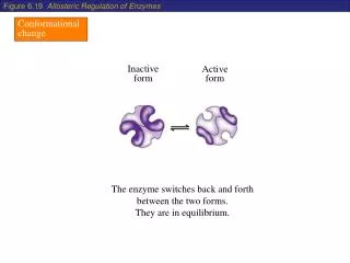
Monod-Wyman-Changeux model • The model makes a number of simple assumptions: • The protein is an oligomer (>1 polypeptide chain) • The protein can exist in 2 states: Tense (T) and Relaxed (R) • T-state has low affinity for oxygen (KT large) • R-state has high affinity for oxygen (KR small) • All the subunits of any one molecule are either in the T-state or the R-state (concerted model)
The Concerted model for allosteric proteins Monod, Wyman and Changeux, MWC model
Koshland, Nemethy, Filmer (sequential) model • Assumes that subunits can change tertiary conformation one at a time in response to binding of substrate. • Cooperativity arises because the presence of some subunits carrying substrate favours the strong binding state in adjacent subunits, whose sites are not yet filled. • As substrate binding progresses almost all the sites become strong binding. • Characterized by existence of molecules with some subunits in weak binding and some in strong binding state.
The sequential model for allosteric proteins Daniel Koshland Increasing O2 binding affinity
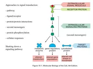
Global regulation by catabolite activation/ repression Diauxic growth High glucose no cAMP CAP does not bind not positive control for dozens of operons Low glucose cAMP is produced CAP binds many operons are activated
Effect of cAMP on CRP and transcription initiation cAMP binds CRP, allosterically changing the conformation of the protein The CRP-cAMP complex binds to target 22 bp DNA Binding of CRP-cAMP complex to the DNA changes the conformation of both the protein and the DNA Altered conformation modulates transcription initiation by RNA polymerase
Structure of the cAMP Receptor Protein HTH motif DNA-binding domain C-helix cAMP-binding domain Kolb et al., Ann Rev Biochem, 1993
34 Å 42 Å Comparison of cAMP+CRP with cAMP+CRP+DNA 1G6N Passneret al., 2000 1O3T Chen et al., 2001
Evidence for cAMP-mediated structural changes in CRP • Free CRP binds DNA with low affinity in absence of cAMP, with high affinity and sequence specificity in the presence of cAMP • CRP is resistant to proteolysis, but is readily digested in the presence of micromolar concentrations of cAMP • DTNB induces intersubunit disulfide bond formation between Cys178s in the CRP dimer in the presence of micromolar concentrations of cAMP but not in its absence • Binding of ANS results in an increase in fluorescence intensity with a shift of emission maximum from 530nm to 480 nm. In the presence of micromolar concentration of cAMP, this fluorescence signal is decreased • Three cAMP analogs: • Induce structural changes and activate transcription • Induce structural changes but do not promote DNA binding or transcription activation • Bind CRP but do not induce structural changes
Crystallographic statistics • Mtb CRP: • Rv3676 • ~22 kDa • P212121 • a=54.1, b=84.6, c= 101.2
DNA binding domain cAMP binding domain Superposition of monomers of Mtb CRP
Grey: cAMP-bound form Blue: cAMP-free form
Helix F Helix E Helix C cAMP Aligned DI
F C LEU 68 LEU 183 LEU 157 ILU 71 mtb 1G6N
Buried surface areas between cAMP-binding and DNA-binding domains
Superposition of Monomers of CRP A chain and B chain Green- DNA bound CRP Grey- cAMPbound CRP Blue- cAMPfree CRP
Helix E: Q170 – G177 and Helix F: S179 – E181 1O3T Difference distance map between cAMP+CRP and cAMP+CRP+DNA structures Helix E: Q170 – G177 and Helix F: S179 – E181 1G6N
Helix E: T168 – G177 and Helix F: S180 – R181 1G6N Helix E: Q177 – V184 and Helix F: R188 – T190 Our Structue Difference distance map between apo CRP and cAMP+CRP structures
3H3U 1G6N
Normal Mode Analysis • Elastic Network Model as implemented in the ElNemo server • Two end- structures: one with cAMP and one without • Homology models of cAMP-free E. coli CRP and cAMP-bound Mtb CRP • Anlayze low frequency normal modes with maximum overlap and collectivity
Dynamic Cross Correlation Map for 1G6N and model of E. coli cAMP-free CRP.
Dynamic Cross Correlation Map for 3H3U and MtbcAMP-bound CRP model.
Normal Mode Analysis of 1G6N and E.colimodel based on 3H3U structure as a reference. Mode 13 shows 57.9 % collectivity and 43.9 % overlap.
NMA of mode 13 shows significant change in C- helix when cAMP binding domain is aligned.
34 Å 45 Å 42 Å 1O3T Mtb CRP 1G6N Comparison of 1O3T , 1G6N and 3H3U
Summary of Overall Conformational Changes Effected by cAMP-binding In absence of cAMP, the cAMP-binding and DNA-binding domains interact closely with each other, reducing mobility of the DNA-binding domain The reduced mobility prevents sequence specific recognition of DNA Binding of cAMP triggers reorientation of side chains (especially Arg 123) in the binding pocket of CRP The cAMP-binding domain is drawn towards the C-helix closing over the bound cAMP Conformational change in the cAMP-binding domain forces out the DNA-binding domain away from the C-helix The DNA-binding domain remains sufficiently flexible, poised for sequence-specific DNA recognition