Download
1 / 40
400 likes | 799 Views
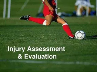
Off-the Field Injury Evaluation. Evaluation of Sports Injuries. Essential skill Four distinct evaluations Pre-participation (prior to start of season) On-the-field assessment Off-the-field evaluation (performed in the clinic/training room…etc) Progress evaluation.

E N D
Evaluation of Sports Injuries • Essential skill • Four distinct evaluations • Pre-participation (prior to start of season) • On-the-field assessment • Off-the-field evaluation (performed in the clinic/training room…etc) • Progress evaluation
Injury Evaluation vs. Diagnosis • While ATC can recognize injury, by law they cannot diagnose --only a doctor can • Doctors of specific regions are allowed to diagnose conditions in those regions (dentist) • Fine line between evaluation and diagnosis • Athletic trainer must act within limits of his/her ability and training and act in accord with professional ethics
Basic Knowledge Requirements • ATC must have general knowledge of anatomy and biomechanics as well as hazards associated with particular sport • Anatomy • Surface anatomy • Key surface landmarks provide examiner with indications of normal or injured structures • Body planes and anatomical directions • Points of reference
Abdominopelvic Quadrants • Four corresponding regions of the abdomen • Divided for evaluative and diagnostic purposes
Musculoskeletal Anatomy • Structural and functional anatomy • Encompasses bony and skeletal musculature • Neural anatomy useful relative to motion, sensation, and pain • Standard Terminology • Used to describe precise location of structures and orientation • Biomechanics (foundation for assessment) • Application of mechanical forces which may stem from within or outside the body to living organisms • Pathomechanics - mechanical forces applied to the body due to structural deviation - leading to faulty alignment (resulting in overuse injuries)
Understanding the Sport More knowledge of sport allows for more inherent knowledge of injuries associated with sport and better injury assessment Descriptive Assessment Terms Etiology - cause of injury or disease Pathology - structural and functional changes associated with injury process Symptoms- perceptible changes in body or function that indicate injury or illness (subjective)
Sign - objective, definitive and obvious indicator for specific condition • Degree- grading for injury/condition • Diagnosis- denotes name of specific condition • Prognosis- prediction of the course of the condition • Syndrome - group of symptoms and signs that together indicate a particular injury or disease
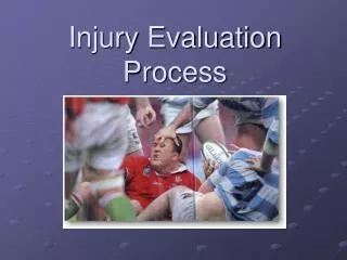
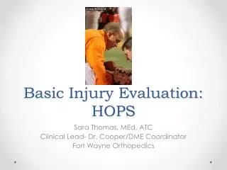
Off-the-field Injury Evaluation • Detailed evaluation on sideline or in clinic/training room setting • May be the evaluation of an acute injury or one several days later following acute injury • Divided into 4 components • HOPS
History Obtain subjective information relative to how injury occurred, extent of injury, MOI Ask the following questions What is the problem? How and when did it occur? Did you hear or feel something? Which direction did the joint move? Characterize the pain
Observations How does the athlete move? Is there a limp? Are movements abnormal? What is the body position? Facial expressions? Asymmetries postural mal-alignments or deformities? Abnormal sounds? Swelling, heat, redness, inflammation, swelling or discoloration?
Palpation Used at the start or further into the evaluation Bony and soft tissue palpation Perform systematically - begin away from the injured site Start with light pressure followed gradually by deeper pressure Bony
Special Tests • Used to detect specific pathologies • Compare inert and contractile tissues and their integrity Active Range of Motion (AROM) • Should be first movement assessment • Assess quality of movement through different ranges and planes at varying speeds and strengths • Pain free throughout full range should be tested while applying force or resistance
Passive Range of Motion(PROM) • Athlete must remain relaxed to remove influence of contractile tissue • Try to classify feel of endpoints Normal • soft tissue approximation- soft, spongy - painless stop • capsular feel-abrupt, hard and firm • bone to bone- distinct abrupt stop • muscular - springy Abnormal • Empty - movement beyond anatomical limits with pain • Spasm - involuntary muscle guarding • Loose - occurs in extreme hypermobility • Springy block - rebound at endpoint • Throughout PROM ATC looking for limitation in movement and presence of pain • Report of pain before end range indicates acute inflammation (stretching and manipulation would be contraindicated) • Pain synchronous with end range indicates subacute and involves inert tissue fibrosis • If no pain at end range, injury is chronic and contractures have replaced inflammation
Resisted Motions (RROM) Evaluate status of contractile tissue Isometric contraction at mid range Different from manual muscle test which occurs throughout ROM Different grading systems used to identify severity and degrees of strength (Cyriax) Goniometric Measurements Measure joint ROM (degrees) Full ROM is major factor in determining return to activity To perform measurement goniometer is placed on lateral aspect of extremity, with 0 or starting position in anatomical positions
Athlete will move either active or passively through available range to endpoint Stationary arm should be placed parallel to long axis of fixed reference part while moveable arm is placed along axis of moveable segment Accuracy and consistency requires practice and repetition Manual Muscle Testing Used to determine vary extent of injury to contractile tissue Limitation in muscular strength is generally caused by pain Generally performed so muscle or group of muscles can be isolated and tested through a full range while applying manual resistance
Ability to move through range or offer resistance is subjectively graded by ATC according to various classification systems Neurological Examination Test 5 major areas (cerebral, cranial nerve, cerebellar, sensory functioning, reflex testing and referred pain) Most musculoskeletal injuries do not require cranial, cerebral or cerebellar assessment and exam can focus on peripheral neurological functioning Cerebral functioning Questions assess general affect, consciousness, intellectual performance, emotional status, sensory interpretation, thought content, and language skills Cranial Nerve function Quality assessed through assessments of smell, eye tracking, facial expressions, biting down, balance, swallowing, tongue protrusion, and shoulder shrug
Cerebellar Function Control of purposeful coordinated movement Touch finger to nose, finger to finger, heel-toe walking Sensory Testing Determine distribution of dermatomes and peripheral nerves Assess Superficial sensation Superficial pain Deep pressure pain Sensitivity to temperature Sensitivity to vibration Position sense
Reflex testing Reflex refers to involuntary response to a stimulus Three types - deep tendon, superficial and pathological Deep tendon reflex (somatic) Caused by stimulation of stretch reflex Biceps (C5) brachioradialis (C6) triceps (C7) patella (L4) Achilles (S1) Superficial reflexes Elicited by stimulation of skin at specific sites producing muscle contraction Upper abdominal (T7,8,9), lower abdominal (T11, 12) cremasteric (S1, 2), gluteal (L4, S3) Absence of reflex = lesion of cerebral cortex Pathological Also superficial reflexes Indicative of lesion in cerebral cortex Babinski’s sign, Chaddock’s, Oppenheim’s, Gordon’s
Determining Projected or Referred Pain Deep burning pain, or ache that is diffuse or in area of no sign of malfunction or disorder is most likely referred Cyriax considers common sites of pain in order of importance - joint, tendon, muscle, ligament, and bursa Pressure on dura mater or nerve sheath can also produce referred pain or sensory response Myofascial trigger points are not related to deep, referred pain (tense tissue bands) Testing Joint Stability A number of specific tests are used to test ligamentous stability for each specific joint Allows clinician to grade severity of injury and determine extent of dysfunction
Testing Accessory Motions The manner in which one articular surface moves relative to another Normal accessory motion must occur to allow for full and un-compromised range of motion Can be impacted by capsular tightness or tightness of musculotendinous units Testing Functional Performance Used to determine athletes readiness to participate or continue participation Used for progress evaluation during rehab Should proceed gradually from relatively easy task to more challenging --mimicking actual sport participation Questions whether athlete has regained full ROM, strength, speed, endurance, and neuromuscular control and is pain free
Postural Examination Many conditions can be attributed to body malalignment Used to look at asymmetries by comparing body relative to grid or plumb line Anthropometric Measurements Science of measuring the body Includes osteometry, craniometry, skin-fold measurements, height and weight. Also involves measurements of limb girth Volumetric Measurements Used to determine changes in limb volume caused by swelling which can be attributed to hemorrhaging, edema or inflammation Measure water that is displaced from a tank in which limb is immersed
Progress Evaluations • When rehab is occurring, follow-up evaluations must be performed to monitor progress • Seeing the athlete daily allows for daily modification • Progress evals should be based on healing process at any given time - providing a framework for the rehabilitation and sometime constraints on progress • Progress evaluations are generally more limited in scope - focus on specific injury and progress relative to previous day • Should still follow similar outline to evaluation
History • Pain comparison (today vs. yesterday) • Movement, better or worse relative to pain? • Treatment - effective or not? • Observations • Degree of swelling • Degree of movement relative to yesterday • Is athlete still guarding? • What is athlete’s affect? Attitude and mood? • Palpation • What is consistency of swelling and has it changed? • Is it still tender to touch? • Deformity compared to yesterday
Special Tests • Do ligamentous tests result in pain and what is the grade? • How do ROM, accessory motion and manual muscle tests compare today to yesterday? • How does the athlete perform in functional tests?
Manual Muscle Testing An evaluation system for diagnosis of disease or dysfunction of the musculo-skeletal and nervous systems
Purpose • Measures the capability of muscles or groups to provide support and movement • Diagnostic tool • Postural balance • Gait impairment • Range of motion • Uses little equipment • Obtains information not defined by other procedures
Precautions • Do No Harm (use gentleness) • Know ROM limits • Follow procedure • Record • Promptly • Accurately
To Get Standardized Results • Proper training and education • Knowledge base of anatomy, physiology and neurology of muscle function • Follow precise testing protocol • Practice, Practice, Practice • A skill developed and maintained with number of cases
Validity and Accuracy • Coordinate the muscle testing findings with other standard diagnostic procedures • The amount of pressure used to test may vary between persons performing the test. • The amount of strength loss must be greater than approximately 20 to 30% to be dependably measurable • Comparison of both sides is a better indicator of loss
Muscles • 3 Types: Skeletal, Smooth, Cardiac • Skeletal around 40% of muscle composition • Generally voluntarily controlled • Composed of fibers • Work in groups • Movement depends on how the muscles are attached
Structure of Muscle Movement depends on how the muscles are attached
How Do Muscles Cause Movement • Origin- where the muscle is attached to the bone; this bone will move very little • Insertion- muscle attachment to bone with most motion • Belly of muscle- part of muscle that enlarges on contraction
Muscle Groups Trapezius Latisimus dorsi Deltoids Biceps Triceps • Quadriceps • Hamstrings • Calf • Low back • Abdominals • Pectoralis major • Rhomboids
Conduct Strength Testing • Correct positioning is essential (Start in extended anatomical position) • Exert uniform force directly on the line opposing movement
Testing of Bicep & Tricep • Support humerus where gravity is against the bicep and tricep, client in anatomical position • Move elbow through full ROM (Passive ROM) • Flexion • Extension • Internal rotation • External rotation
Maneuver to Assess Muscle Strength • With arm in full extension, pull downward on forearm while client attempts to flex. • With arm flexed, apply pressure against forearm, ask client to straighten arm. • When performing muscle tests, be sure to evaluate for asymmetry of the muscle groups (i.e. atrophy on one side and not the other) and landmarks prior to testing.
Use the following scale to rate strength: • 0-No movement, no contraction of the muscle • 1- Trace, evidence of muscle contraction but no joint movement • 2- Poor, complete range of motion with gravity eliminated • 3-Fair, complete range of motion against gravity • 4- Good, complete range of motion against gravity with moderate resistance • 5-Normal, complete range of motion against gravity with maximal resistance without evidence of fatigue
Other Test Results • Weakness – defined as a strength below fair in non weight bearing muscles; below fair + in weight bearing muscles • Contracture – degree of shortness in muscle, so it cannot move through ROM • Substitution – weak muscles are supported by other muscles to move
Strength Test Examples • Gastrocnemius • Rectus Femoris • Brachioradialis