Download
1 / 41
580 likes | 3.12k Views
Coryneform bacteria, listeria and erysipelothrix. Corynebacterium sp. Listeria monocytogenes Erysipelothrix rhusiopathiae. Corynebacteria. Significant Corynebacterium species C. xerosis C. pseudodiphtheriticum C. pseudotuberculosis C. jekeium C. ulcerans Rhodococcus equi

E N D
Coryneform bacteria, listeria and erysipelothrix Corynebacterium sp. Listeriamonocytogenes Erysipelothrixrhusiopathiae
Corynebacteria • Significant Corynebacterium species • C. xerosis • C. pseudodiphtheriticum • C. pseudotuberculosis • C. jekeium • C. ulcerans • Rhodococcus equi • Arcanobacterium haemolyticum
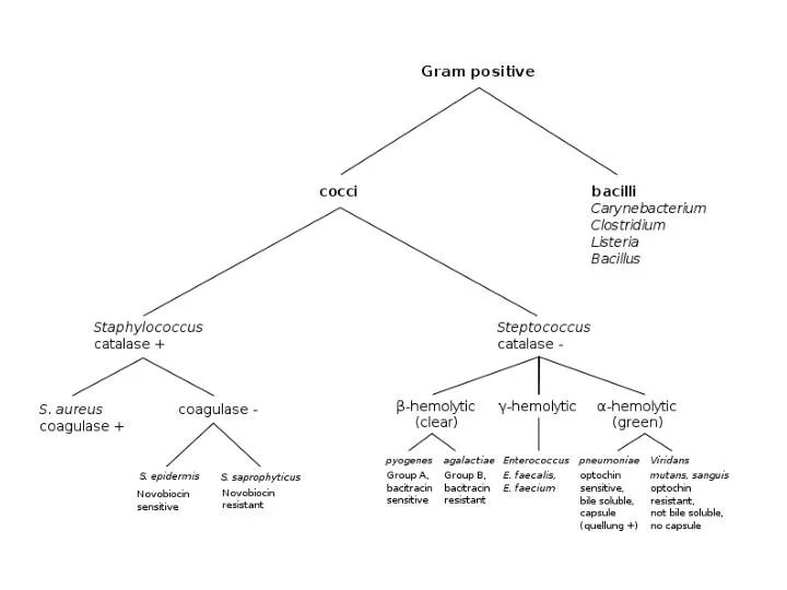
Corynebacterium Species • General characteristics • Found as free-living saprophytes in fresh and salt water, in soil and in the air • Members of the usual flora of humans and animals(often dismissed as contaminants) • Often called “diphtheroids” • Corynebacterium diphtheriae isthe most significant pathogen • Other species may cause infections in the immunocompromised hosts
Corynebacterium Species:General Characteristics • Morphology • Gram-positive, non–spore-forming rods • Arrange in palisades:“L-V” shape; “Chinese characters” • Pleomorphic: “club-ends” or coryneform • Beaded, irregular staining
C. diphtheriae: Agent of Diphtheria • Toxigenic Corynebacterium diphtheriae • Worldwide distribution but rare in places where vaccination programs exist • Exotoxin, Diphtheria toxin, as the virulence factor • Not all C. diphtheriae strains produce toxin • Toxin is produced by certain strains • Toxin is antigenic
Toxigenic Corynebacterium diphtheriae • Toxin consists of two fragments • A: Active fragment • Inhibits protein synthesis • Leads to cell/tissue death • B: Binding • Binds to specific cell membrane receptors • Mediates entry of fragment A into cytoplasm of host cell
Clinical Forms of Diphtheria • Respiratory • Acquired by droplet spray or hand to mouth contact • Non-immunized individuals are susceptible • Non-respiratory • Systemic • Skin and cutaneous forms
C. diphtheriae: Causative Agent of Diphtheria • Respiratory disease–diphtheria • Incubation period–2 to 5 days • Symptoms: sore throat, fever, malaise • Toxin is produced locally, usually in the pharynx or tonsils • Toxin causes tissue necrosis, can be absorbed to produce systemic effects • Forms a tough grey to white pseudomembrane which may cause suffocation
C. diphtheriae: Causative Agent of Diphtheria • C. diphtheriae pseudomembrane • WBC + organism
Clinical Infections: Non-Respiratory Disease • Systemic infections • Toxin is absorbed in the blood stream and carried systemically • Affects the kidneys, heart, and nervous system • Death occurs due to cardiac failure • Cutaneous form • More prevalent in the tropics • Infections occur at the site of minor abrasions • Maybe superinfected with Streptococcus pyogenes and/or Staphylococcus aureus
Treatment • Infected patients treated with anti-toxin and antibiotics • Anti-toxin produced in horses • Antibiotics have no effect on circulating toxin, but prevent spread of the toxin • Penicillin drug of choice
Laboratory Diagnosis • Microscopic morphology • Gram-positive, non–spore-forming rods, club-shaped, can be beaded • Appear in palisades and give "Chinese letter" arrangement • Produce metachromatic granules or “Babes’ Ernst” bodies (food reserves) which stain more darkly than remainder of organism Corynebacterium diphtheriae gram stain
Laboratory Diagnosis:Cultural Characteristics • Loeffler's slant or Pai's slant—Used to demonstrate pleomorphism and metachromatic granules ("Babes’ Ernst bodies“) • Growth on Serum Tellurite or modified Tinsdale exhibits brown or grayish→ to black halos around the colonies Tellurite: tellurium dioxide (TeO2).
Laboratory Diagnosis:Cultural Characteristics • Blood agar plae (BAP) 24-48 hours at 37oC small, grey translucent colonies • Small zone of b- hemolysis also s een
Laboratory Diagnosis • Identification • Confirm identification by fermentation reactions(glucose +) • Catalase positive • Urease negative • Non-motile
Laboratory Diagnosis • Toxigenicity testing • Elek test • Immunodiffusion test • Organisms are streaked on media with low Fe content to maximize toxin production. • Identification of C.diphtheriae does NOT mean the patient has dipheria. Must show the isolate produces the toxin. protease peptone agar + serum (horse or bovine) 1 and 4 positive
C. diphtheriae • Treatment: antitoxin • Prevention: DPT immunization
C. jekeium • Clinical Infections • Septicemia • Meningitis • Pulmonary disease • Populations Affected • Immunosuppressed • IV drug users
C. jekeiumColony Morphology • Isolation & identification • BAP: 48-72 hours at 35oC in ambient air or 5% CO2 small, gray-white colony, nonhemolytic • Gram stain: pleomorphic, club-shaped gram positive rod arranged in V forms or palisades
C. jekeiumLab Diagnosis • Identification • Nitrate reduction= negative • Urea= negative • Sucrose= negative • Glucose= positive
C. jekeium • Susceptibility testing • Exhibits resistance to multiple antibiotics • Susceptible to vancomycin
Listeria monocytogenes:General Characteristics • Gram-positive, non–spore-forming rods • Only human pathogen in genus • Widespread in nature • Known to infect a wide variety of animals • Human exposure is limited; direct or indirect • Transient colonization occurs without disease
Listeriamonocytogenes:Clinical Infections • Adults • Septicemia/meningitis in the compromised/elderly • Mild flu-like syndrome in pregnant women could be fatal to fetus • Ingestion of contaminated food • Neonatal • Early onset from intrauterine transmission results in sepsis; high mortality rate • Late onset manifests as meningitis; lower mortality rate
Listeria monocytogenes: • Virulence Factors • Hemolysin ( Listeriolysin O)- damages macrophage • Catalase • Superoxide dismutase • Phospholipid C • P60 surface protein- induces phagocytosis thru adhesion and penetration
Laboratory Diagnosis: L. monocytogenes • Identification • Microscopic morphology • Gram Positive non–spore-forming coccobacillary, pairs or short chains • Colony Morphology • Grows well on blood agar; colonies produce a narrow zone of hemolysis similar to Group B Streptococcus • Small, round and translucent
Laboratory Diagnosis: L. monocytogenes • Grows well at 0.5° C to 45° C • Because of this temperature range, especially the cooler end of the range, this organism grows well in refrigerated products, such as cream, cheese, deli meats, etc. • Can sometimes be isolated after “cold enrichment” (hold broth at 4° C for several weeks and subculture)
Laboratory Diagnosis: L. monocytogenes • Identification • Catalase positive • Motility: • Motile at 25o C; "umbrella" type → • Tumbling motility in hanging drop preparations (this can be seen on Gram Stain Tutor at www.medtraining.org) “Umbrella” motility pattern (Left) typical for L. monocytogenes
Laboratory Diagnosis: L. monocytogenes • Identification • CAMP test • Produces a “block” type of hemolysis in contrast to “arrow”-shape produced by Group B Streptococcus CAMP test with Listeria monocytogenes Positive CAMP test for Group B Streptococcus
Differentiating Characteristics betweenL. monocytogenes and Other Gram Positive Bacteria
Erysipelothrix rhusiopathiae:General characteristics • Gram positive, non–spore-forming, pleomorphic rods (can produce long filaments) • Distributed in nature • Can cause disease in animals (swine, turkey, sheep); swine is the main reservoir • Humans acquire the infection through occupational exposure, such as cuts & scratches (fish handlers, animal products)
Erysipelothrix rhusiopathiae:Clinical Infections • Erysipeloid • Self-limiting localized infection at the site of inoculation • Produces painful swelling, usually on the hands or fingers • Heals within 3 to 4 weeks • Endocarditis • May occur in those who have had valve replacements • Disseminated infections may occur, but rarely
Laboratory Diagnosis:Erysipelothrix rhusiopathiae • Microscopic Morphology • Pleomorphic, gram-positive thin rods that may form long filaments, may be arranged singly, in short chains, or in a V shape
Laboratory Diagnosis: Erysipelothrix rhusiopathiae • Identification • Catalase negative • CO2 is required • Distinguishing characteristic: Production of H2S on TSI • Microaerophilic • Nonmotile • Test tube brush growth in semisolid motility media
Laboratory Diagnosis: Erysipelothrix rhusiopathiae • Colony Morphology • Grows on blood or chocolate agar—colonies may appear gray or translucent, pinpoint with alpha hemolysis or nonhemolytic
Treatment: Erysipelothrix rhusiopathiae • Penicillin, cephalosporin, erythromycin
Characteristics of Corynebacterium, Listeria, and Erysipelothrix
Lactobacillus • Widely distributed in nature • Normal flora of mouth, GI tract and female genital tract • Treat with pencillin plus an aminoglycoside • resistant to vancomycin (helps in diagnosis) • Clinical Infections • Bacterial vaginosis • Bacteremia, endocarditis, meningitis (rare)
Lactobacillus • Microscopic Morphology • Long, slender gram positive pleomorphic bacilli • Non-spore forming • Colony Morphology • Microaerophilic • SBA: pinpoint, α- hemolytic colonies • Lab Diagnosis • Catalase negative