Download

1 / 64
640 likes | 755 Views
Clinical Staging in the Multidisciplinary Management of the Cancer Patient. Frederick L. Greene, M.D. Department of Surgery. Today: Carolinas Medical Center Carolinas HealthCare System. Pierre Denoix. american joint committee on cancer.

E N D
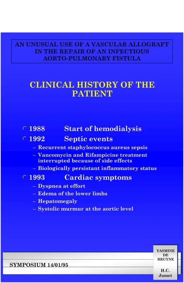
Clinical Staging in the Multidisciplinary Management of the Cancer Patient Frederick L. Greene, M.D.Department of Surgery
IT IS THE BEST THING , IN MY OPINION, FOR THE PHYSICIAN TO APPLY HIMSELF DILIGENTLY TO THE ART OF FOREKNOWING Hippocrates(400BCE)
Staging • Provides a framework for discussion • Helps stratify patients into groups that are prognostically and therapeutically similar • Facilitates comparison across large populations
Biologic Progression of Tumor • Tumor first grows locally • Metastasizes to regional lymph nodes • Finally, metastasizes systemically
Prognostic Factor Variable that can explain some of the heterogeneity associated with the expected course and outcome of a disease. from Tannock & Hill. The Basic Science of Oncology, 1998
Criteria for Prognostic Factors I. Statistically significant - prognostic value only rarely occurs by chance II. Independent - retains prognostic value when combined with other factors III. Clinically relevant - has a major impact on prognostic accuracy
Application of Prognostic Factors Clinical practice • Select appropriate diagnostic tests • Select optimal treatment plan • Predict the outcome for an individual patient • Select appropriate follow-up monitoring • Patient and caregiver education
Tumor Related Prognostic Factors • Pathology • morphology, grade, growth pattern, • Anatomic tumor extent • TNM, tumor bulk, number, tumor markers • Tumor biology • proliferation indices, molecular markers, genetic markers • Symptoms • Performance status
Approximately 80 prognostic variables are reported for breast cancer.
Traditional Prognostic Parameters for Human Mammary Carcinoma Tumor FactorsHost Factors Lymph node status Age Tumor size Menopausal status Histologic/nuclear grade Familial history Lymphatic/vascular invasion Previous neoplastic disease Pathologic stage (TNM) Immunosuppression Steroid receptor status (ER/PR) Host inflammatory response DNA content (ploidy, S-phase) Nutrition EIC (in situ) Prior chemotherapy Prior radiation
AJCC - Changing Strategies of TNM Staging Summary: Breast • Axillary nodes (routine H&E or IHC) • N1 1 to 3 • N2 4 to 9 • N3 10+ • Infraclavicular nodes are N3 • Supraclavicular nodes reclassified as N3
Molecular Prognostic Factors Prediction of occult disease • Need for adjuvant therapy • Prediction of natural history • Patient survival • Prediction of treatment response • Overlap of factors
Staging and Tumor-Related Prognostic Factors • Cancer-related proteins • Tumor markers, PSA, CEA • Gene products • Tumor suppression, ras, src, c-myc, p53, pRB • Tumor-cell apoptosis • bc1-2/bax, caspase family
Staging and Tumor-Related Prognostic Factors • Cancer-related proteins • Cell-cycle control • p53, cyclins, Ki67 • Metastasis and angiogenesis • CD 44, VEGF, angiogenesis inhibitors
Patient Related Prognostic Factors • Demographics • age, race, gender, level of education, socioeconomic status, religion • Co-morbidity • fixed - inherited conditions • changeable - weight, coexistent illness, cardiac and renal function • Performance status • Compliance
Prognostic Factors - Environment Treatment • Physician focus • Quality and accuracy of diagnosis, • Expertise, timeliness of action • Health care system focus • Access to care, diagnostics, screening • Society focus • Socioeconomic status, insurance status • Distance from treatment center
Residual Tumor Classification • Strongest predictor of outcome • Reflects effects of therapy • Influences further therapeutic procedures • Importance of identification by surgeon • Histologic confirmation of gross residual tumor
Category I:Well supported by the literature, generally used in patient management and of sufficient importance to modify TNM stage group • RX Presence of residual tumor cannot be assessed • RO No residual tumor • R1 Microscopic residual tumor • R2 Macroscopic residual tumor Residual Tumor (R)
Prognostic Factors TNM + R + Prognostic Factors
Isolated Tumor Cells (ITC) (single tumor cells or small clusters) (≤ 0.2 mm) VS Micrometastases ( 0.2 cm in greatest dimension)
AJCC - Changing Strategies of TNM Staging Breast - Additional Descriptors “sn” suffix: Based only on sentinel lymph node dissection without an axillary node dissection eg. pN0(sn) or pN1(sn)
Isolated Tumor Cells and Micrometastasis pNO: No regional lymph node metastasis histologically, no examination for isolated tumor cells (ITC) pNO(i-): No regional lymph node metastasis histologically, negative morphologic findings for ITC pNO(i+): No regional lymph node metastasis histologically, positive morphologic findings for ITC
Isolated Tumor Cells and Micrometastasis pNO(mol-): No regional lymph node metastasis histologically, negative nonmorphologic findings for ITC pNO(mol+): No regional lymph node metastasis histologically, positive nonmorphologic findings for ITC
National Cancer Data Base (NCDB) • A community-based oncology management and outcomes data base • A joint project: ACS/ACoS
NCDB: Strengths • Provides information on patterns of care throughout the United States • Able to evaluate both rare and common malignancies • Only data source with ability to provide feedback at local level where care is delivered • Low cost
NCDBClinical Surveillance • Diagnosis / AJCC Stage • Treatment by AJCC Stage • Patient Demographics • Survival by Treatment & Stage
Elements Absent from NCDB • Co-morbidity • Provider data • Delivery system • Complications • Patient Satisfaction • QOL
NCDB Annual Data Collection • Current diagnosis/accession year • 5-year follow-up cases • 10-year follow-up cases • 15-year follow-up cases
Study Population • 50,042 patients (node-positive) • Study period - 1987 through 1993 • Hospitals reporting - 1,705 (28%)
STAGING of COLON CANCER(Greene et al,Ann Surg2002;236,416-21) Stage III (T1-4, N1-2, MO) Stage III A (10.7%) Stage III B (60.5%) Stage IIIC (28.8%) T1/2, N1 T3/4, N1 Any T, N2
5-Year Observed Survival Rates: Stage III Colon Cancers by AJCC 6th Edition Subgroup, Cases Diagnosed 1987-1993
AJCC 7th Edition • Available early October 2009 • Effective with cases diagnosed on or afterJanuary 1, 2010 • Editions • 6th ends on December 31, 2009 • 7th begins January 1, 2010
Change • “There is nothing more difficult to carry out, nor more doubtful of success, nor more dangerous to manage, than to initiate a new order of things.” • Machiavelli
TNM of H & N Cancer The terms “Resectable” and “Unresectable” replaced with “Moderately Advanced” (Resectable) “Very advanced” (Unresectable) Descriptor added to N staging Extracapsular spread (ECS) ECS + or ECS – Does not influence nodal staging system
Regional Lymph Nodes-H&N Prognostic importance of nodal involvement Survival significantly worse Based on location Nodes beyond first echelon of nodal drainage Particularly nodes in lower regions of neck Designate if nodes above or belowlower border of cricoid Cartilage
Summary of Changes-Thyroid T1 has been subdivided T1a Tumor ≤ 1cm limited to the thyroid T1b Tumor >1 to ≤ 2cm limited to the thyroid Changed descriptors for T category s solitary tumor m multifocal tumor Terminology changed for T4 tumors. T4a. Moderately Advanced (replaced Resectable) T4b.Very Advanced (replaced Unresectable)
Primary Tumor (T4)* T4a Moderately advanced diseaseTumor of any size with major extrathyroid extension (beyond the thyroid capsule to invade subcutaneous soft tissues, larynx, trachea, esophagus, or recurrent laryngeal nerve) T4b Very advanced diseaseTumor invades prevertebral fascia or encases carotid artery or mediastinal vessels *All Anaplastic carcinomas are staged T4
Strategies for the 7th Edition of TNM • Utilization of SEER data base->109,000 patients • Division of T4 into T4a and T4b • T4a-penetrates visceral peritoneum • T4b-Tumor directly invades other organs or structures • IIA-T3N0 • IIB- T4aN0 ; IIC-T4bN0
Highlights of Changes - Breast • T • Guidance on determining tumor size • Clarification of inflammatory carcinoma • Recommend grading with Nottingham • N • Classification of isolated tumor cells is more stringent • Restricted use of (sn) modifier to 5 or fewer nodes • M • Created new cM0 (i+) category • Disseminated tumor cells detectable in bone marrow • Circulating tumor cells • Incidental in other tissues < 0.2 mm
Melanoma Photo courtesy of Kelly McMasters
T CLASSIFICATION:Diagnosis after January 1, 2010 Thickness T1: <1.0 mm T2: 1.1-2.0 mm T3: 2.1-4.0 mm T4: >4.0 mm Ulceration/Mitoses T1 T1a: no ulcer and mitoses < 1/mm2 T1b: ulceration present or mitoses > 1/mm2 Ulceration T2-4 T2a – no ulcer T2b – with ulcer…