Download
1 / 65
650 likes | 1.07k Views
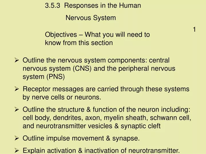
3.5.3 Responses in the Human Nervous System. 1. Objectives – What you will need to know from this section. Outline the nervous system components: central nervous system (CNS) and the peripheral nervous system (PNS)

E N D
3.5.3 Responses in the Human Nervous System 1 Objectives – What you will need to know from this section • Outline the nervous system components: central nervous system (CNS) and the peripheral nervous system (PNS) • Receptor messages are carried through these systems by nerve cells or neurons. • Outline the structure & function of the neuron including:cell body, dendrites, axon, myelin sheath, schwann cell, and neurotransmitter vesicles& synaptic cleft • Outline impulse movement & synapse. • Explain activation & inactivation of neurotransmitter.
2 • The structure and function of a neuron: variation in size and shape. • Neuron -- Three part structure:> dendrite(s) receive information and carry it towards thecellbody,> the axon conducts nerve impulses away from the cell body,> the cell body contains the nucleus and other organelles andproduces neurotransmitter chemicals. • Explain the role & position of 3 types of neuron -- sensory/motor/inter • Movement of nerve impulse.(Detailed knowledge of electrochemistry not required.) • Knowledge that the conduction of nerve impulses along a neuron involves movement of ions (details not required).
3 • Outline the senses with the brain as an interpreting centre. • Outline the CNS, brain & spinal cord. • State location & function of cerebrum / hypothalamus / pituitary gland / cerebellum / medulla oblongata • Label &/or draw diagrams of spinal cord (cross section) indicating : white matter, grey matter, central canal, 3 layer protective tissue-meninges. • Spinal nerves containing dorsal and ventral roots that project from the spinal cord
4 • Outline disorders from NS disorders: paralysis/Parkinson's including: Cause/Prevention/Treatment • Outline PNS including the location nerve fibres & cell bodies. • State the role, structure & mechanism of the Reflex arc/action. • The sense organs contain receptors, with the brain as an interpreting centre for received information. • Knowledge of the five senses and related organs. • Study the eye and the ear – recognition and fuction of the main parts. • Corrective measures for long and short sight or for a hearing defect.
The Nervous System • Organisms must be aware of what is happening around them,as this affects their survival chances. • Co-ordination of an organism’s activities is carried out by thenervous system and the endocrine system. • A nervous system allows an organism to detect and respond to stimuli in its internal or external environment. • A stimulus is any change in your environment e.g. a flash of light, a noise, a fly landing on your nose.
The nervous system relies on electrical signals, carried byspecialised cells [neurons], and is involved in fastresponses. • The central nervous system(CNS) is your brain and spinalcord. • The brain keeps a check on internal organs and activities, such as the level of carbon dioxide orwater in theblood. • The peripheral nervous system (PNS) is the nerves branchingfrom the CNS to all parts of the body.
Endocrine Cells involved Gland Message Chemical (Hormone) Carried by Blood Message sent to Cells throughout the body Received by Target organ Comparison of the endocrine and nervous systems - 1
Endocrine Speed of transmission Usually slow Effects Can bewidespread Duration long-lasting (hours) Comparison of the endocrine and nervous systems -- 2
LEARNING CHECK • Co-ordination of an organism’s activities is carried out by what 2 body systems? • Distinguish between the CNS and PNS. • What is a neuron? • What is an impulse? • List the main differences between the endocrine and nervous systems.
NS ----- Co-ordination & Response • A stimulus is any change in your environment • A receptor is a nerve cell that detects the stimulus • A neuron is a specialised cell that carries electrical messages(impulses) around the body. • An impulse is an electrical message that is carried along aneuron.
Neuron structure • The cell body contains the nucleus and other organelles andproduces neurotransmitter chemicals. • A dendron is a short fibre that receives information and carries ittowards the cell body. • The axon is a very long fibre that conducts impulses away from the cell body.
A dendron is a short fibre that receives information and carry ittowards the cell body. • Dendrites are small branches of a dendron. • Many Schwann cells wrap their fatty cell membranes around an axon, forming a myelin sheath. • The myelin sheath insulates the neuron from electrical impulses flowing in other neurons.
There are three types of Neuron : • Sensory neuron –carries messages from the sense organ tothe central nervous system (CNS). • Interneuron –connects sensory and motor neurons and socarries messages within the CNS. • Motor neuron –carries impulses from the CNS to muscles andglands.
SYNAPSE • Synapse—region where two neurons come into close contact. • Synaptic cleft—the gap between two neurons, bridged bychemicals (neurotransmitters). • Neurotransmitter—chemical released across a synaptic cleft tocarry a signal from one neuron to another. • The chemical is then destroyed or removed
LEARNING CHECK • What is a stimulus? • What is a receptor? • Name the main parts of a neuron and give one function of each. • Distinguish between a sensory and motor neuron. • Distinguish between synapse and synaptic cleft. • What are neurotransmitters?
The Brain • The brain is composed of over 100 billion neurons, eachreceiving messages simultaneously from thousands of otherneurons. • The brain is protected by the skull bones, meninges (threemembranes) and cerebrospinal fluid.
The cerebrum is our conscious brain, with different parts havingdifferent jobs to do. cerebrum hypothalamus pituitary • The hypothalmus is the centre for the regulation of the internalorgans. • The pituitary ‘[master] gland secretes hormones that stimulateother glands to release their hormones.
4) The brain decides to move away the hand 3) Here another sensory neuron carries the signal to the brain 5) This impulse is sent by MOTOR NEURONS to the hand muscles (the effectors) via the spinal chord… 2) The impulse is carried by SENSORY NEURONS to the spinal chord 1) Receptors in your skin detect a stimulus 6) Which then moves the hand away Stimulus Receptor Sensory NeuronCoordinator Motor Neuron Effector Response Conscious actions A conscious action is one where the brain makes a considered response. Here’s what happens:
Medulla oblongata • The cerebellum co-ordinates processes that we have learnedto do automatically, such as speaking. cerebellum • The medulla oblongata co-ordinates involuntary, automaticprocesses—such as breathing, heartbeat.
LEARNING CHECK • Name the 3 main parts of the brain and one function of each. • How is the brain protected? • What is meant by “conscious action”? • What does the term “involuntary” mean? • Distinguish between the cerebrum and the cerebellum.
The spinal cord is well protected by the vertebrae, meninges(three membranes) and cerebrospinal fluid. • It transmits impulses to and from the brain and controls manyreflex actions.
A cross-section through the spinal cord shows a small central canal, filled with cerebrospinal fluid, surrounded by an area of grey matter, shaped somewhat like the letter H.
Grey matter contains cell bodies and dendrites (regions of a neuron that have no white myelin covering). • Outside the grey matter, the spinal cord consists of whitematter (containing axons only).
In humans, 31 pairs of spinal nerves branch off from the spinal cord. • Each spinal nerve has a dorsal root and a ventralroot. • The dorsal root consists of nerve fibres carrying information into the spinal cord fromthesenses. • The dorsal root ganglion is a swelling that consists of the cell bodies of the sensory neurons.
The ventral root consists of nerve fibres carrying information out from the spinal cord, to the muscles and glands. • The cell bodies of the motor neurons are positioned within the grey matter of the cord. • The spinal cord transmits impulses to and from the brain and controls manyreflex actions.
Interneuron REFLEX ACTION --- The Reflex Arc • A reflex action is a quick, automatic response to a particularstimulus.
Interneuron REFLEX ACTION --- The Reflex Arc Suppose you touch a hot flame. • Almost instantly you pull your hand away. • In this brief instant, a message has been carried by a sensoryneuron from pain receptors in the skin to the spinal cord.
Interneuron • In the spinal cord, the message is passed on to an interneuron and then to a motor neuron, and so into muscles that respond by contracting and pulling your hand from the flame.
Interneuron • This response saves the body frominjury. • The response is called a reflex action, as it does not involve conscious control, and is predictable andautomatic. • Many of the activities of the body, such as breathing and keeping our balance, are regulated by reflex actions.
LEARNING CHECK • How is the spinal cord protected? • Distinguish between grey and white matter. • Distinguish between dorsal and ventral root • What is meant by reflex action? • Give some examples of reflex action> • What is an interneuron? • Distinguish between cell bodies and ganglions.
Nervous System Disorder • Parkinson’s disease is a nervous system disorder, normally seen in older people, in which muscles become rigid and movement is slow and difficult, with persistent tremors [shaking]. • It is caused by the brain reducing the normal amount of dopamine that it makes. • There is at present no means of preventing it, but giving L-dopa (which the body changes into dopamine) can relieve the symptoms in many patients.
SENSE ORGANS • Animals have specialised senses to provide them with information about their environment. • The five senses are sight, hearing, touch, taste and smell. • A receptor is a cell that can detect a stimulus • A stimulus is any change in your environment, e.g. light, sound.
Sense Sense Sense Organ Organ Stimulus detected Sight Sight Sight Eye Eye light [by rods and cones in the retina] Hearing Hearing Hearing Ear Ear sound [receptors in cochlea] Touch Touch Touch Skin Skin touch, pressure, temperature and pain [receptors spread throughout body] Taste Taste Taste Tongue Tongue chemicals [taste buds detect sweet, sour, salt and bitter]. Smell Smell Smell Nose Nose chemicals [receptors in the nasal cavity detect vapours]
Eyelid The EYE • Eyelids can cover and protect the eyes. Conjunctiva Cornea • Conjunctiva—thin transparent lining protecting the cornea. • Cornea—front transparent part of the sclera. It focuses light rays on the retina.
Sclera • Sclera—tough fibrous outer layer – the ‘white’ of the eye; it maintains the shape of the eyeball. Choroid Retina • Choroid—contains blood vessels supplying food and oxygen to the cells of the eye. • Retina—the innermost layer that contains the receptor cells [rods and cones].
Fovea • The fovea is where our best vision is [mainly cones] • The front region of the choroid is specialised into the iris Iris • Iris—contains blood vessels and melanin [giving us our eye colour], and controls the amount of light entering the eye [through the pupil].
Pupil • In bright light, pupil constricts. • In dim light, the pupil dilates.
Ciliary body [muscle]—thickened edge of the choroid that controls the shape of the lens Ciliary muscle • Suspensory ligaments—hold the lens in place. Suspensory ligaments Lens • Lens—like a magnifying glass, it focuses the light rays on the retina.
Lens—focuses the light rays on the retina. • Accommodation is the ability of the lens to change its shape (focal length) to form a clear image.
LEARNING CHECK • Name the 5 senses and the organs involved. • Name the 3 main layers of the eye and the function of each. • What is the function of the [a] iris, [b] lens, [c] cornea, [d] fovea • What is accommodation?
Close Vision • For close vision, the ciliary muscle contracts, the suspensory ligaments relax, the lens becomes thicker.
Distant Vision • When the eye is at rest, the lens is thin, has a long focal length and is adapted for seeing distant objects.
Accommodation is the ability of the lens to change its shape (focal length) to form a clear image.
Seeing things at different distances For distant objects, the ciliary muscle relaxes and so the suspensory ligaments pull tight,pulling the lens thinner – the light doesn’t bend as much. For close objects the ciliary muscle contracts, allowing the lens to go fat, thus bending the light more.
Aqueous humour—watery liquid that supplies the lens and cornea with nutrients and helps keep the shape of the cornea and lens. Aqueous humour Vitreous humour • Vitreous humour—gel that helps maintain the shape of the eye.