Download
1 / 24
240 likes | 343 Views
Anaphase. Prophase. Metaphase. INTERPHASE G1 phase . Metabolic changes prepare the cell for division. At a certain point - the restriction point - the cell is committed to division and moves into the S phase.

E N D
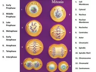
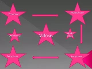
Anaphase Prophase Metaphase
INTERPHASE G1 phase. Metabolic changes prepare the cell for division. At a certain point - the restriction point - the cell is committed to division and moves into the S phase. S phase. DNA synthesis replicates the genetic material. Each chromosome now consists of two sister chromatids. G2 phase. Metabolic changes assemble the cytoplasmic materials necessary for mitosis and cytokinesis. M phase. A nuclear division (mitosis) followed by a cell division (cytokinesis). The period between mitotic divisions - that is, G1, S and G2 - is known as interphase
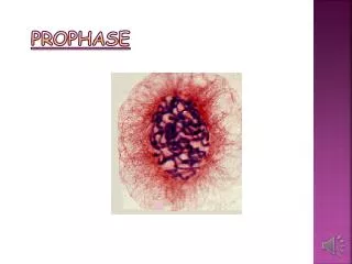
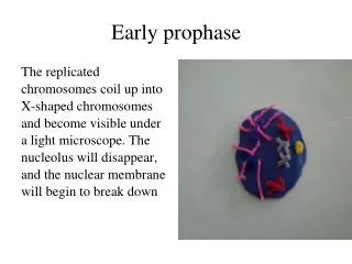
Prophase Prophase occupies over half of mitosis. The nuclear membrane breaks down to form a number of small vesicles and the nucleolus disintegrates. A structure known as the centrosome duplicates itself to form two daughter centrosomes that migrate to opposite ends of the cell. The centrosomes organise the production of microtubules that form the spindle fibres that constitute the mitotic spindle. The chromosomes condense into compact structures. Each replicated chromosome can now be seen to consist of two identical chromatids (or sister chromatids) held together by a structure known as the centromere.
METAPHASE • The chromosomes, led by their centromeres, migrate to the equatorial plane in the midline of cell - at right-angles to the axis formed by the centrosomes. • This region of the mitotic spindle is known as the metaphase plate. • The spindle fibres bind to a structure associated with the centromere of each chromosome called a kinetochore. Individual spindle fibres bind to a kinetochore structure on each side of the centromere. • The chromosomes continue to condense. • The chromosomes align themselves along the metaphase plate of the spindle apparatus.
ANAPHASE • The shortest stage of mitosis. • The centromeres divide, and the sister chromatids of each chromosome are pulled apart - or 'disjoin' - and move to the opposite ends of the cell, pulled by spindle fibres attached to the kinetochore regions. • The separated sister chromatids are now referred to as daughter chromosomes. (It is the alignment and separation in metaphase and anaphase that is important in ensuring that each daughter cell receives a copy of every chromosome.)
TELOPHASE • The final stage of mitosis, and a reversal of many of the processes observed during prophase. • The nuclear membrane reforms around the chromosomes grouped at either pole of the cell, the chromosomes uncoil and become diffuse, and the spindle fibres disappear.
The final cellular division to form two new cells. In plants a cell plate forms along the line of the metaphase plate; in animals there is a constriction of the cytoplasm. The cell then enters interphase - the interval between mitotic divisions
Explain how mitosis ensures that daughter nuclei are genetically identical.