Download
1 / 8
80 likes | 158 Views
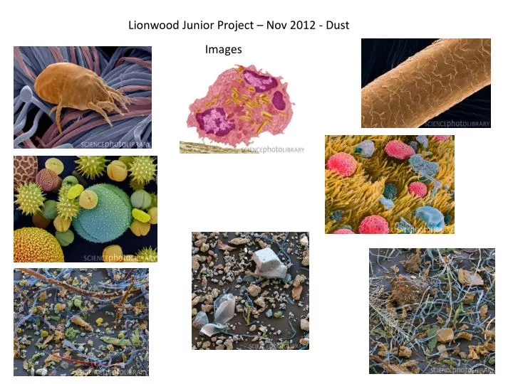
Lionwood Junior Project – Nov 2012 - Dust. Images. Dust mite, SEM - Z445/0328 Power and Syred , SPL

E N D
Dust mite, SEM - Z445/0328 Power and Syred, SPL Caption: Dust mite. Coloured scanning electron micrograph (SEM) of a dust mite (Dermatophagoides sp.) on the threads of a piece of fabric. The mite's head is at lower right. Millions of dust mites inhabit the home, feeding on shed skin cells. They mainly live in furniture, and are usually harmless. However, their excrement and dead bodies may cause allergic reactions in susceptible people. Magnification: x1725 at 6x7cm size. All images from Science photo library – no permission to use as yet! Pollen grains, SEM - C012/4970 AMI Images, SPL Caption: Pollen grains. Coloured scanning electron micrographs (SEM) of pollen grains from a variety of plants. Including: sunflower (Helianthus annuus), morning glory (Ipomeapurpurea), hollyhock (Sildalceamalviflora), lily (Liliumauratum), primrose (Oenotherafruticosa), and caster bean (Ricinuscommunis). Allergens in trachea - M320/0156 Eddy Gray, SPL Caption: Trachea covered in allergens. Coloured Scanning Electron Micrograph (SEM) of the surface of the trachea (windpipe) with breathed in pollen and dust. These airborne particles may cause asthma or hay fever (allergic rhinitis). Pollen grains are coloured pink. The surface of the trachea is made up of cells with hair-like cilia (yellow). Together with mucus, these cilia serve to trap airborne particles and by beating upwards in a wave-like motion they remove foreign matter from the air tubes and lungs. In asthmatic or allergic patients, such particles may lead to a hyper- sensitive reaction causing breathing difficulties. Magnification: x2,000 at 6x7cm size. x6,500 at 8x10" House dust - living room, SEM - C011/5072 Eye of science, SPL Caption: House dust. Coloured scanning electron micrograph (SEM) of a sample of dust from a living room shelf. This sample contains: cotton fibres (blue), plant matter (green), animal material (dander, beige), pollen (yellow), mineral particles (grey), various fibres (red), and hair (brown). Magnification: x107 when printed 10 centimetres wide. Eosinophil white blood cell, TEM - C014/1437 Steve Gschmeissner, SPL Caption: Eosinophil white blood cell. Transmission electron micrograph (TEM) of a section through an eosinophil. Eosinophils, like all white blood cells, are part of the body's immune system. They are involved in the body's allergic response and help to defend the body from invading parasites. The cell's cytoplasm contains many characteristic granules (yellow) that contain enzymes used to destroy invading organisms. Magnification: x5000 when printed 10 centimetres wide. House dust - bedroom, SEM - C011/5074 Eye of Science, SPL Caption: House dust. Coloured scanning electron micrograph (SEM) of a sample of dust from a bedroom. This sample contains: a spring (white), cotton fibres (blue), hair (brown), plant matter (green), animal material (dander, beige), pollen (reddish), and soil (brown). Magnification: x60 when printed 10 centimetres wide. House dust - kitchen, SEM - C011/5073 Eye of science, SPL Caption: House dust. Coloured scanning electron micrograph (SEM) of a sample of dust from a kitchen. This sample contains: a sugar crystal (upper right), the wing of an insect (lower left), cotton fibres (blue), flour (beige), starch grains (white), and other particles of plant origin. Magnification: x70 when printed 10 centimetres wide. Human hair SEM - C014/0306 Pascal Goetgheluck, SPL Caption: Human hair. Coloured scanning electron micrograph (SEM) of a strand of human hair. Hairs are made up of dead tissue. The outside of the hair, the cuticle, is covered in overlapping scales that protect the hair's central cortex, which is made up of the fibrous protein keratin. Flakes of dead skin, SEM - C001/1720 Steve Gschmeissner, SPL Caption: Flakes of dead skin. Coloured scanning electron micrograph (SEM) of dead skin from the scalp (dandruff), a condition where there is an excessive shedding of dead skin cells. Dandruff can be the result of psoriasis, a fungal infection, or the overproduction of sebum, the secretion from sebaceous glands associated with hair follicles. Magnification: x35 when printed at 10 centimetres wide.
Science session – dust detectives! P1210394.jpg SAM_0948.jpg SAM_0957.jpg P1210382.jpg SAM_0951.jpg P1210385.jpg
Writing SAM_0969.jpg Historian, Samuel O’Rourke P1210390.jpg P1210389.jpg Dustmite poem .jpg
Art SAM_0962.jpg P1210400.jpg P1210408.jpg P1210404.jpg P1210406.jpg P1210402.jpg
Dustmite habitat quiz to build the scene.. SAM_0974.jpg SAM_0971.jpg SAM_0975.jpg P1210417crop.jpg P1210413.jpg
School display SAM_1033.jpg