Download
1 / 30
340 likes | 888 Views
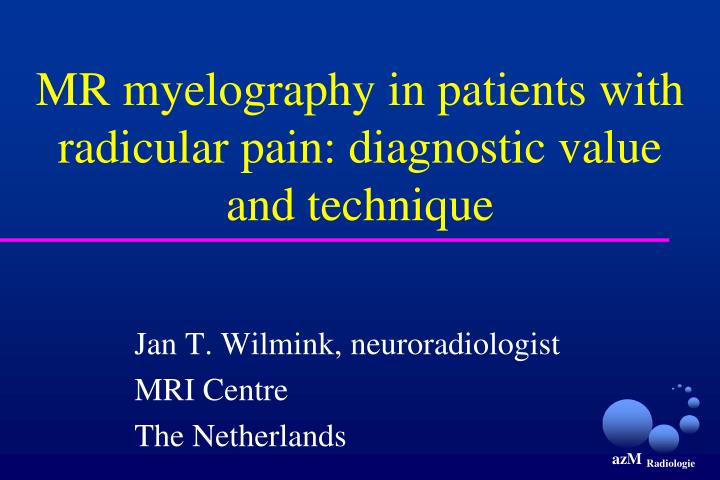
MR myelography in patients with radicular pain: diagnostic value and technique. Jan T. Wilmink, neuroradiologist MRI Centre The Netherlands. Problem: clinical relevance of MRI findings. 98 asymptomatic, 27 symptomatic subjects. L4-L5: protrusion. L5-S1 extrusion.

E N D
MR myelography in patients with radicular pain: diagnostic value and technique Jan T. Wilmink, neuroradiologist MRI Centre The Netherlands
L4-L5: protrusion L5-S1 extrusion
protruded discs are frequently asymptomaticextruded discs usually cause symptoms(???) So:
Problem: rating scales assessing only disk displacement fail to take into account size of spinal canal
L5-S1 extrusion S1 root L5-S1 extrusion
So: let’s think root:myelography radiculography caudography
T2W MR myelogram X-ray myelogram
Sequence for T2 FSE MR myelography- heavy T2 weighting TR/TE 6000/450 - slice thickness 4mm, overcontiguous- echo train length 65- FOV small: 150mm- MIP postprocessing to produce virtual image of dural sac- long acquisition time 6:30mins per projection
multishot 6:30min single shot 1.5sec
multishot single shot
single shot x 10 32.5sec multishot 6:30min
Single-shot single-slice T2 FSE MR myelography with multiple excitations- heavy T2 weighting TR/TE 6500/1270, 10 excitations - single slice, thickness 30mm, oblique x2, no MIP needed - echo train length 256- scan matrix 256, reconstruction matrix 512- scan percentage 75- FOV 150mm, rectangular 75%- acquisition time 32.5secper projection, total 65sec
1. Patient with left sciatica Illustrative cases
midsagittal left lateral T1W SE sagittal
upper disc level lower disc level herniation, no root compression herniation, root compression?? T1W axial, L4-L5
L5 root compressed normal S1 root and root sleeve
2. Patient with backache irradiating to left buttock Illustrative cases
root compression?? !! !!
3. Patient with backache and some irradiation to both legs Illustrative cases
? left L5-S1 extrusion, S1 root compression?
in 43 patients MR myelography reduces diagnostic uncertainty from 19 cases to 6 cases
Conclusions- MR myelography (MRM)is valuable add-on but cannot replace standard MR examination - MRM is useful in cases when disc lesion is seen but effect on root is uncertain (compressed or not)- MRM findings must always be matched against standard MR images and clinical presentation- with acquisition time of only 1-2 mins, the MRM sequence should be included in standard spinal study