Download
1 / 32
380 likes | 880 Views
MASS ANALYZERS AND IONIZATION METHODS. Greg Barrett-wilt , phd UW Mass spectrometry/proteomics facility. Central components of a mass spectrometer. Ionization source. Mass analyzer/filter. Detector. Vacuum chamber. T o perform mass spectrometry two things are required:

E N D
MASS ANALYZERS AND IONIZATION METHODS Greg Barrett-wilt, phd UW Mass spectrometry/proteomics facility
Central components of a mass spectrometer Ionization source Mass analyzer/filter Detector Vacuum chamber To perform mass spectrometry two things are required: 1. The analyte must be an ion 2. The analyte must be in the gas phase
Electron impact ionization (EI) “Hard” ionization method – ionization and fragmentation occur simultaneously Incompatible with liquid streams Widely used with gas chromatography Very standardized method: 70eV electron energy NIST database
Electron impact ionization (EI) http://www.chromacademy.com/essential-guide/nov2010/fig-1.jpg Wiley Registry of 638,000 compounds (>700,000 spectra) NIST Library: 242,477 compounds Fred McLafferty
Chemical ionization (CI) “Softer” ionization method – observation of intact molecular ions possible EI source filled with gas (N2, methane, ammonia) yields ionized gas molecules Reagent gas ions ionize analytes Primary ion formation CH4 + e-→ CH4+ + 2e- Secondary reagent ions CH4 + CH4+ → CH5+ + CH3 CH4 + CH3+ → C2H5+ + H2 Product ion formation M + CH5+ → CH4 + [M+H]+ (protonation) [M+H]+
matrix assisted laser desorption ionization (MALDI ) “Soft” ionization method: preserves the intact analyte UV absorbing matrix is energized by the laser Matrix molecules desorbed from the surface carry analyte molecules into the gas phase Proton transfer from matrix (acid) to analyte in the gas phase UV laser used to generate ions: N2 (337 nm) or Nd:YAG (355nm) Yields singly-charged species almost exclusively http://www.magnet.fsu.edu/education/tutorials/tools/ionization_maldi.html Koichi Tanaka: Nobel Prize in Chemistry (2002)
maldi MALDI matrices α-cyano 4 hydroxycinnamic acid (CHCA) 3-hydroxy picolinic acid (3-HPA) dihydroxy benzoic acid (DHB) sinapic acid Analyte samples are co-crystalized with matrix molecules Example from AB Sciex 4800 TOF-TOF spotted with CHCA
MALDI Couples well to high mass-range instruments (TOF) because high molecular weight biomolecules with a single charge will be observed at high m/z. [M+H]+ BSA protein standard [M+2H]2+ [2M+H]+ [3M+2H]2+ [3M+H]+ [4M+H]+
Electrospray ionization (ESI) “Soft” ionization that yields intact analyte ions with one or more charges A high voltage (kV) is applied to a liquid stream Flow rates can vary between ~50nL/min and 1mL/min These various flow rates require substantially different source parameters John Fenn: Nobel Prize in Chemistry (2002)
Electrospray ionization (ESI) Couples easily to HPLC Analyte molecules can accept multiple charges (protons) Analytes can be present in multiple charge states 432.899 100 +3 Peptide DRVYIHPFHL (MW=1295.677) +33 Relative Abundance +32 +31 Protein MW=31,928 +30 +4 +2 +29 324.926 648.844 +28 0 300 350 400 450 500 550 600 650 700 750 800 850 900 950 1000 m/z
Quadrupole V = AC voltage (a) http://www.nasa.gov/mission_pages/msl/news/sam-tastes-mars.html (q) Ubiquitous mass filters for numerous applications Randall Pedder “Practical Quadrupole Theory: Graphical Theory (2010) (Presented as ASMS poster 2001)
Quadrupole Mass range: 10 - ~2000 Resolution: “unit” (100 at m/z 100, 1000 at m/z 1000) Sensitivity: moderate-low (scanning instrument) Spectral acquisition rate: moderate (~1s per spectrum) Implications for proteomics: Full MS only, low resolution, slow speed
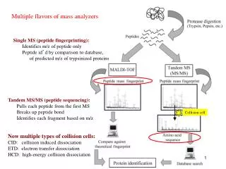
Triple-Quadrupole Tandem mass spectrometer: two stages of mass filtering (MS/MS) Collision cell between two quadrupole mass filters: Collision induced dissociation (CID) Additional experiments possible with a triple quadrupole SRM/MRM Domon and Aebersold, Science312 (2006)
Triple-Quadrupole Triple quadrupole instruments are a very active field of mass spec development. Considered the most widely-used type of mass spectrometer. Agilent 6495 (2014) AB Sciex 6500 (2012) Thermo TSQ Quantiva (2013)
Triple-Quadrupole General operating specifications same as single-quadrupole Mass range: 10 - ~2000 Resolution: “unit” (100 at m/z 100, 1000 at m/z 1000) Sensitivity: moderate-low (scanning instrument) Spectral acquisition rate: moderate (~1s per spectrum) BUT in SRM/MRM mode: Mass range: N/A Resolution: “unit” (~1 amu mass window) Sensitivity: very high (fixed mass, no scanning) Acquisition rage: N/A (~10ms per transition) Implications for proteomics: Very high sensitivity in SRM/MRM mode, very linear instrument response, excellent quantitation
3D ion trap 3D ion trap introduced commercially in 1983 Substantial increase in full scan sensitivity because all m/z ions are trapped and detected (as opposed to quadrupole instruments). Very efficient MS/MS, especially for peptides. Can only trap a limited number of ions at one time. Wolfgang Paul and Hans Dehmelt: Nobel Prize in Physics (1989) V= ring electrode voltage amplitude = ring electrode RF frequency r0= trap radius
2D ion trap (linear ion trap) “Can only trap a limited number of ions at one time.” 3D ion trap: ~1000 ions in a spherical volume 2D ion trap: ~30,000 ions in a cylindical volume Thermo LTQ: 2D trap replaces a 3D ion trap (single mass filter) OR 2D trap incorporated in a hybrid MS (see below) AB Sciex QTRAP: 2D trap replaces Q3 of a triple quad (tandem mass filters) Implications for proteomics: Low resolution, very good peptide MS/MS
Orbitrap Originally described in 1920 (Kingdon trap) it was only in the late ’90’s that it was developed into a mass spectrometer (Makarov, Anal. Chem. 2000). First new mass filter since the quadrupole ion trap in 1984 Technology owned by ThermoFisher Commercial instrument introduced in 2005 Resolving power = >200,000 http://en.wikipedia.org/wiki/Orbitrap#cite_note-Mak1-1 m/z analysis property:
LTQ Orbitrap Operation Principle 1. Ions are stored in the Linear Trap 2. …. are axially ejected 3. …. and trapped in the C-trap 4. …. they are squeezed into a small cloud and injected into the Orbitrap 5. …. where they are electrostatically trapped, while rotating around the central electrode and performing axial oscillation The oscillating ions induce an image current into the two outer halves of the orbitrap, which can be detected using a differential amplifier Ions of only one mass generate a sine wave signal
Frequencies and Masses The axial oscillation frequency follows the formula Where w = oscillation frequency k = instrumental constant m/z = …. well, we have seen this before Many ions in the Orbitrap generate a complex signal whose frequencies are determined using a Fourier Transformation
Orbitrap Critical feature of Orbitrap hybrid instrument: Multiple ion traps means that ions can be analyzed simultaneously in the different analyzers. R = 120,000 Significant increase in duty cycle (neither mass analyzer is ever idling) http://www.youtube.com/watch?v=KjUQYuy3msA Implications for proteomics: High resolution/mass accuracy (Orbitrap), very good peptide MS/MS (ion trap), very high duty cycle (hybrid), fast scan speed
Orbitrap XL: LC/MSn 100 ~1000 proteins in 4 hours 90 80 70 60 Relative Abundance 50 40 30 20 10 0 40 60 80 100 120 140 160 180 200 220 240 260 280 Time (min)
Several instrument types Orbitrap Elite (2010) Q Exactive (~2008) LTQ Orbitrap XL (~2007) Orbitrap Fusion (2013)
Time-of-flight (TOF) Where k incorporates accelerating voltage and distance of flight tube
Time-of-flight: maldi AB Sciex 4800 MALDI TOF-TOF: MS and MS/MS
Q-tof (or Qq-TOF) Similar to triple-quad MS, but the third quad is a TOF High resolution, accurate mass MS/MS is performed after MS survey scan (in series) by increasing the offset energy in the collision cell High-resolution MS1 and MS/MS Assist in database searching of proteomic data, unknown i.d.
Ion mobility MS Agilent 6560 Adds a drift tube in the flight path of the ions prior to the TOF region This allows for separation of species based on their gas-phase cross-section: Small ions are retarded by gas molecules less than large ions (even for same m/z) Waters
TOF features MALDI TOF: Decoupled from HPLC: fast (~1 sec/spectrum) MS/MS capable (TOF/TOF): ideal for rapid protein ID from 1D gel band Resolution > 20,000 Laser rate 1kHz-2kHz (new instruments) ESI-TOF: Resolution >40,000 Fast electronics give excellent sampling across chromatographic peaks Q-TOF: High resolution MS1 and MS/MS Ion Mobility Q-TOF: Additional structural information from cross-section (separate isobarics) Non-covalent interaction experiments Implications for proteomics: High resolution/accurate mass for MS and MS/MS, fast scan speed, wide mass range (100,000 MS1, 4000 MS/MS)
Wrap-up Ionization sources: EI CI MALDI ESI Mass Analyzers: Single and triple quadrupole 3D and 2D ion trap Orbitrap Time-of-flight (TOF/TOF) Q-TOF Ion mobility Others: APCI APPI ICP SIMS Others: Magnetic sector Isotope ratio FT-ICR Accelerator