Download
1 / 14
440 likes | 1.43k Views
Laser Eye Surgery. And other surgical vision correction. Requisite Drawing of the Eye. Sclera – The tough white material that makes up the wall of the eye Cornea – The clear portion of the sclera that allows light to pass into the eye and aids in focusing

E N D
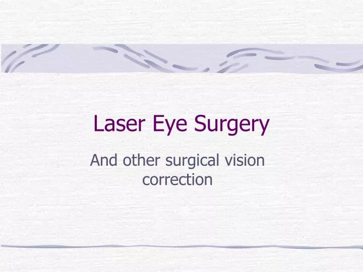
Laser Eye Surgery And other surgical vision correction
Requisite Drawing of the Eye • Sclera – The tough white material that makes up the wall of the eye • Cornea – The clear portion of the sclera that allows light to pass into the eye and aids in focusing • Lens – The crystalline lens though which light passes in order to reach further depths of the eye. Also aid in focusing light • Retina – The inner layer of the eye, a sheet of neural tissue which contains the receptors which kick off perception
Problems Corrected By Surgery Myopia Hyperopia Astigmatism
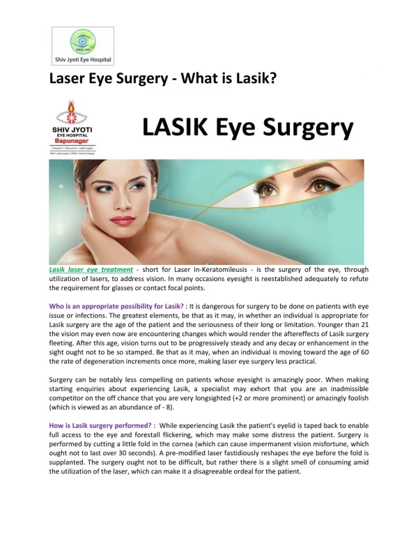
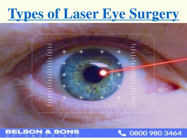
LASIK • Laser-Assisted In Situ Keratomileusis • Uses a knife, called a microkeratome, to cut a flap in the cornea and an eximer laser to reshape the exposed stroma (the middle layer of the cornea) • Keratomileusis, a predecessor • to LASIK, involved removing • a section of the cornea, • reshaping it, and then • replacing it
Procedure • Contact lenses change the shape of the cornea for up to several weeks after they’re worn. Glasses, therefore, must be worn for 2-4 weeks before the initial visit as well as in the time before surgery. • The contours of each eye are mapped out in a computerized topographical analysis, to provide a detailed plan for what will be removed during surgery. The thickness of the cornea will also be measured.
Before surgery, analgesic drops will be administered to numb the eye. The area around the eye will be cleaned, and a speculum will be inserted to keep the eye open. • A ring will be applied to create suction to the cornea. The microkeratome is then attached to the ring and a flap is cut into the cornea. • The ring and microkeratome are removed and the flap is folded back to expose the sclera. The laser is then positioned over the eye, whereupon the patient must fixate upon a red light. • Pulses of energy will destroy the pre-selected areas of tissue. Any debris are then washed from the eye, and the flap is returned to its original position. • A clear shield is placed over the eye, and the patient is then allowed to leave, returning for check-ups 1 day, 1 week, 2 weeks, 3 months, 6 months, and 1 year after surgery
Afterwards • For the first few months after surgery, visual acuity will fluctuate • Vision will stabilize in 3-6 months, but during that period of stabilization glares, halos, and difficulty driving at night may persist • If touch-ups are needed, they should be done in 3 months time, when the vision is fairly stabilized and a new flap need not be cut. Instead, the old one can simply be pried up. If done later, the progress of the healing will require a new flap to be cut
Possible Side Effects • Glare, halo, and difficulty driving at night, if they remain after the stabilization period, will be a permanent feature, and will not be improved by re-operation • Corneal thinning, which if extreme can lead to more serious issues, can result in artificially low tonometry (intra-ocular pressure) readings • May be under-treated or over-treated • Vision loss, or bad vision in low contrast situations • Severe dry eye syndrome • Results may diminish with age, and natural effects of ageing on the eye will still occur
PRK • Photorefractive Keratotomy • A laser is used to reshape the surface of the cornea, removing the outer layer of the cornea in the process. A protective lens is placed over the eye to protect for the 3-5 days it takes for the outer layer to regrow. Because no corneal flap is created, the procedure is easier to perform and less delicate afterwards, but more uncomfortable for the patient
RK • Radial Keratotomy • Slits are cut in the eye to correct myopia (decreasing curvature) or astigmatism (rounding the football shape)
Other Procedures • Intracorneal Implants • Laser Thermal Keratoplasty • Refractive Lens Exchange
Bibliography • Food and Drug Administration. <http://www.fda.gov/> • The Physician and Sports Medicine. <http://www.physsportsmed.com/> • <http://www.avclinic.com/> • <http://www.eyesearch.com/> • <http://www.emedicine.com/> • <http://www.foreyesight.com/> • American Academy of Ophthalmology. <http://www.aao.org/>