Download
1 / 46
590 likes | 1.07k Views
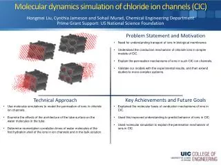
Chloride Channels. Yuanting Lu Cristian Escobar. 09/23/2011.

E N D
Chloride Channels Yuanting Lu Cristian Escobar 09/23/2011
Cells actively transport Cl- across the plasma membrane by transporters that accumulate Cl- intracellularly (Cl- loaders including the Na+-K+-Cl- cotransporters NKCC, Cl--HCO3- exchangers AE, and Na+-Cl- cotransporters NCC) or pump Cl- out of the cell (K+-Cl- cotransporters KCC and Na+-dependent Cl- - HCO3- exchanger NDCBE). Cl- flows passively across a variety of Cl- channels in the plasma membrane including Ca-activated Cl- channels (CaCC), cAMP-activated Cl- channels (CFTR), cell-volume regulated anion channels (VRAC), and ligand-gated anion channels (GABAA and glycine receptors). In addition, Cl- channels and transporters are found in intracellular membranes, such as the endosomallysosomal pathway, and play a role in regulating intra-vesicular pH and Cl- concentration. Intravesicular pH and [Cl-] are important in vesicular trafficking.
Functions • Stabilization of membrane potential. • Depolarization • Cell volume regulation. • Fluid transport in epithelia. • Neutralization of H+ in lysosomes. ClC-1 ClC-2 GABA Glycine Bestrophin TMEM16A VDCC ClC-Kb CFTR ClC-3 Voltage dependent Ligand gated Cl- channels Ca2+ Volume sensitive Cl- channels pH cAMP pH
CLC Chloride Channels Stabilization of membrane potential Influx of Cl- suppresses depolarizing inputs and stabilizes membrane potential Myotonia and muscle fiber type changes. Mutations in Na1 (1) or Cl2 (2) channels or the lack of the sarcolemmal chloride channel (ClC-1; 2) can lead to overexcitability of the sarcolemma (myotonia). Normal muscle responds with single action potentials upon single stimuli, whereas myotonic muscle often responds with runs of action potentials. Increased membrane excitation can cause protein kinase C (PKC) activation in the nucleus and changes in the pattern of myogenic regulating factors (MRF). The myogenic factors control gene transcription and therewith couple membrane excitation to the muscle fiber type. A second signaling pathway involves cytoplasmic Ca21. The propagation of action potentials into the transverse tubule system (TT) activates the L-type Ca21 channel and stimulates Ca21 release from the sarcoplasmic reticulum (SR) via the ryanodine receptor (RyR). A Ca21 signaling pathway into the nucleus is suggested.
CLC Chloride Channels Functions Endosomal acidification • H+ concentration inside of lysosomes require Cl- influx to neutralize positive charges. • Some ClC channels mediate Cl−:H+ exchange rather than voltage-dependent anion channel activity.
CLC Chloride Channels Epithelial transport • ClC-Kb transport Cl- acumulated by NKCC trasporter to the intertisium. Fluid transport and Cl– channel. Ascending loop of Henle (thick ascending limb, TAL) is an important part of the tubular segment (left panel), playing a cardinal role in construction of high osmolality in the renal medulla by unidirectional transport of NaCl. Luminal Na+, K+ and Cl– are transported via the Na/K/2Cl transporter into cells without water. Na+ and Cl– are transferred into the basolateral space and thereby concentrated in osmolality. K+ is back-leaked by K+ channels resulting in lumen-positive (compare with interstitium) transtubular potential. This lumen-positive potential drives cations, Ca2+ or Mg2+, through the cell junction (claudin) under the electrochemical gradient. Thus, all 3 modes of transport, Na/K/2Cl transporter, K channel and Cl channel (ClCK) are cardinal in the construction of the high osmolality.
Cystic fibrosis transmembrane conductance regulator (CFTR) • Localization on apical membranes • Plays an important role in fluid secretion in airways, intestine, sweat glands. • Their activity is regulated by cAMP-dependent phosphorylation Model showing proposed domain structure of cystic fibrosis transmembrane conductance regulator (CFTR). MSD, membrane-spanning domain; NBD, nucleotide-binding domain; R, regulatory domain; PKA, cAMP-dependent protein kinase. current-voltage relationship of channel at 357C with symmetrical 140 mM N-methyl-D-glucamine chloride (n 5 for each data point).
CFTR, example of transport Intestine Fig. 2. A model of secretory epithelial cell and secretory diarrhea. Cholera toxin or heat-stable enterotoxin can increase the intracellular cAMP or cGMP levels by activating the membrane-localized adenylate cyclase (AC) or guanylate cyclase (GC). An increase in the intracellular cAMP or cGMP leads to the phosphorylation of the R-domain of CFTR by PKA or cGMP dependent protein kinase II (cGKII), which, in turn, activates the CFTR chloride channel, resulting in Cl secretion into the lumen. As a consequence, Na+ and water are effuxed into the lumen through the paracellular transport mechanism. Therefore, the net result is the secretion of fluid and lectrolytes across the apical surface into the gut lumen. Cl is taken up from the basolateral (blood) side by the Na+–K+–2Cl cotransporter (NKCC). K+ recycles through basolateral K+ channels, and Na+ is pumped out of the cell by the Na+–K+-ATPase.
CFTR, example of transport Airways Cell models for electrolyte transport in human airways. CFTR is the dominant pathway for luminal Cl− exit in human airways, while TMEM16A (ANO1) is probably the major secretory pathway in mouse airways. (ADE: adenosine).
Ca2+ Activated Chloride Channels Regulation of membrane potential by CaCCs
Ca2+ Activated Chloride Channels Functions Physiological roles of CaCCs. In epithelial cells, activation of CaCCs by intracellular Ca2þ elevation leads to Cl secretion followed by transepithelial transport of Naþ and water. In smooth muscle cells, activation of CaCCs is part of an amplification echanism. Intracellular Ca2þ increase by extracellular stimuli activates CaCCs and Cl efflux. The resulting membrane depolarization opens voltage-dependent Ca2þ channels that cause a further intracellular Ca2þ increase, thus potentiating contraction. Another amplification mechanism occurs in olfactory receptors, where the initial Ca2þ increase is triggered by cAMP-gated channels. CaCC is also involved in phototransduction and regulation of neuronal excitability
Ca2+ Activated Chloride Channels TMEM16A (ANO1) Putative topology of TMEM16A channels. TMEM16A (NM 018043) has eight putative transmembrane domains and a p-loop between transmembrane (TM) domain 5 and 6 but no apparent similarity to other ion channels. A number of consensus sites for phosphorylation by kinases are present in the N-terminus, but no binding site for Ca2+ (numbering of amino acids according to splice product a,c,d [13]). Three cysteines (at position 651, 656, 661) are located in the pore forming loop and are accessible to sulfhydryl reagents. ANO1 is activated by intracellular Ca2+ in a voltage-dependent manner. Dose–response relationship of ANO1 activation by Ca2+. Current responses were normalized versus those observed at 10 mM (circles, 260 mV, n58) or 3 mM Ca2+ (triangles, 160 mV, n58). d, An amplitude histogram of single-channel currents activated by Ca2+ at 160 mV.
Ca2+ Activated Chloride Channels Airways Contribution of Ca2+-dependent Cl− secretion by TMEM16A (ANO1) in mouse and human airways. Cell models for electrolyte transport in mouse airways. CFTR is the dominant pathway for luminal Cl− exit in human airways, while TMEM16A (ANO1) is probably the major secretory pathway in mouse airways. (ADE: adenosine).
Ligand Activated Chloride Channels Inhibitory Glycine Receptor • Glycine is a neurotransmitter in inhibitory synapses • GlyR are pentamers of α and subunits (2 α : 3 ). Schematic drawing of the pentameric arrangement of GlyR subunits in homo-oligomeric a1 (left) and heterooligomeric a1b (right) GlyRs. Binding sites for glycine are indicated in yellow, and glycine sites also capable of binding strychnine are shown by a red surround
Ligand Activated Chloride Channels GABA Receptor • 2 types of receptor: • GABAa= LGCC • GABAb= G-protein couple
Volume sensitive Cl- channels • They have been difficult to identify. • MCLC intracelular Cl- Channel Mechanism of cell swelling and a possible role of VDCC in tubulo-glomerular feedback. (A) Mechanism of cell volume regulation and activation of VDCC is shown. The cell under the aniso-osmotic surrounding is illustrated at the top of the panel. The tonicity of extracellular and intracellular solution is expressed on the bottom. Cells swell in hypotonic (indicated as a white column) extracellular solution by an influx of water via aquaporin channels. The influx of water per se or change of volume induces several lines of signal transduction, which then activate Cl– (VDCC) and K+ channels. Efflux of these ions lowers the concentration of ions in the cell interior reaching equilibrium to extracellular tonicity, and then cell volume is restored.
Two key properties for ion channels: • Selective ion conduction • Gating • ClC channels: “fast gating”
Crystal Structure of the EcClCFab complex FIG.1 A Red: Light Fab chain Orange: Heavy Fab chain Blue: ClC subunit Green: ClC subunit Each subunit within the dimer forms its own independent pore for Cl- ions.
EcCLC Crystal structure Carboxyl group FIG. 1.A Scen Selectivity filter Sint FIG. 2.B FIG. 1.B
Whole-cell oocyte currents expressing ClC-0 from Torpedo ray +80 mV to -180 mV Probability to be open WT -100 mV E166 mutants Single channel measurement
Mutant EcClC crystal structure WT E148A E148Q Close Open FIG. 2.B FIG. 4.B FIG. 4.D
Proposed gating mechanism FIG. 5
Conclusions: • When the Sext site is occupied by the glutamate carboxyl group, the pore is closed; When the site is occupied by a Cl- ion, the pore is open. • Two pores are gated independently. • The conformational change is local.
Ca2+ - activated Cl- currents are dispensable for olfaction G.M. Billing, B. Pal, P. Fidzinski & T. J. Jentsch Nature Neuroscience, vol 14(6), 2011
Main olfactory epithelium(MOE) & the vomeronasal organ (VNO) http://www.neuro.fsu.edu/~mmered/vomer/
Olfactory System • Olfactory bulb • Mitral cells • Bone • Nasal epithelium • Glomerulus • Olfactory receptor cells http://en.wikipedia.org/wiki/Olfactory_system
OSN signal transduction pathway The disruption of Ano2 caused no change in G-protein, adenylate cyclase. odorant Ano2 Ca2+ activated Cl- channels CNG cation channels
Ano2 in MOE and VNO FIG. 1
Ano2 in MOE and VNO • Ano2 co-localized with the cilia marker protein ac tubulin in MOE and the Cnga2 subunit of the olfactory CNG channel. FIG. 2
Olfaction is not grossly impaired in Ano2-/-mice Tyrosin hydroxylase helps convert tyrosine to dopamine / norepinephrine /epinephrine.
Ca2+ activated Cl- currentsare absent from Ano2-/- neurons FIG. 5
Disruption of Ano2 moderately reduces electro-olfactograms(EOGs) Grey: NFA Black:Normal Ringer
Conclusions • Disruption of Ano2 in mice virtually abolished Ca2+ -activated Cl- currents in MOE and VNO. • Disruption of Ano2 reduced fluid-phase EOG by only ~40%, did not change air-phase EOG. • Ca2+ -activated Cl- currents are dispensable for olfaction.
Experimental system • Colon Carcinoma epithelial cell line T84 • SPQ (6-methoxy-N-(3-sulfopropyl)quinolinium) • Fluorescence spectrometer configuration:
SPQ emission and excitation spectra SPQ loaded T84 cells Ex Sample 10 µM SPQ in water Ex Sample 443 nm 320 nm 443 nm Em 344 nm Em 344 nm
Stern-Volmer constants F0/F = 1 + Ksv [Q] 2.5 µM SPQ in water SPQ loaded T84 cells SPQ • Nigericin K+/H+antiporter • TributyltinCl-/OH-antiporter • KCl and fluorescein gradient • Fluorescein as internal standar • (ex=495 nm em = 519 nm) Fluorescein
Functional Assay on T84 cell cAMP IBMX isopreterenol Control Glibenclamide (100 µM) CFTR(inh)-172 (5 µM)
Conclusions: • Using SPQ as Cl- sensing molecule can be applied to measure it accurately • This study present an easy method to study CFTR activity.
Thank you !!