Download
1 / 42
420 likes | 846 Views
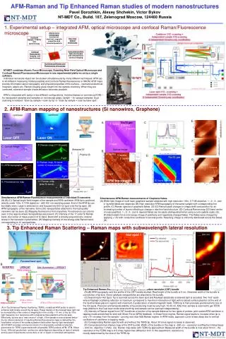
Surface Enhanced Raman Nanotags for Ultrasensitive and Multiplexed Detection of Caner. Ximei Qian and Shuming Nie Emory Univ. Dept. of Biomedical Engineering Atlanta, GA 30322. For SNM, Feb 1st, 2010. Overview. Brief background of SERS spectroscopy

E N D
Surface Enhanced Raman Nanotags for Ultrasensitive and Multiplexed Detection of Caner Ximei Qian and Shuming Nie Emory Univ. Dept. of Biomedical Engineering Atlanta, GA 30322 For SNM, Feb 1st, 2010
Overview • Brief background of SERS spectroscopy • Comparing three optical tagging particles: • Organic chromophores • Semiconductor quantum dots • SERS nanotags (Au nanoparticles) • Evolution of SERS nanotags • First generation (Porter group; Anal. Chem. 1999, 71(21), 4903-4908) • Second generation (Glass-coated tags; Anal. Chem. 2003, 75(22), 6171-6176) • Third generation (Polymer-coated tags; Nature Biotech. 2008, 26(1), 83-90) • Biomedical application of SERS nanotags • In vitro cellular tagging — cancer biomarker detection • In vivo non-invasive transcutaneous tumor detection
Noble Metal Nanoparticles Surface plasmon resonance + + + + + + + + Electrons undergo collective oscillation.
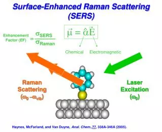
SERS enhanced sensitivity 1014-1016 fold (single molecule detection) Raman Scattering Rayleigh Scattering Surface Enhanced Raman Scattering 1 1014 1014-1016 Sensitivity While most photons are elastically scattered, 1 in 107 incident photons undergo the Raman effect.
Key components of SERS nanotags Core: Au nanoparticle provides EM enhancement for SERS Reporter molecule: provides fingerprint signature; chemical enhancement for SERS Thin-layer protection: reduces non-specific binding/aggregation Function group: for specific targeting
Comparison of SERS tags, QDs and dye molecules SERS tag (60nm) QD705 Atto610 dye • Molecular weight(g/mol) ~109 ~106491 N = fV/Va N: number of gold atom in one nanoparticle f: FCC packing density 0.74 V: volume of one nanoparticle Va: volume of one gold atom a: radius of gold atom 1.44Å
10nm 15nm QD Core Size comparison Dye molecules SERS tags QDs Calculated ~1nm QD705 Au ~57nm 50 nm 50 nm C-C bond length = 1.44Å Volume ratio 107 1 104
SERS tag Atto610 dye QD705 • Molecular weight ~109 ~106491 • Core size 57nm10-15nm1nm • Hydrodynamic size 78±11nm18±6nm
QD705 Atto610 dye SERS tag • Molecular weight ~109 ~106491 • Core size 57nm10-15nm 1nm • Hydrodynamic size 78±11nm18±6nm • Bandwidth 2nm63nm 37nm • Structural information fingerprintbroad structureless • Multiplex detection >5 with NIR excitationlimited limited
After 1 min. Photo-stability
Photo-stability QD705
QD705 Atto610 dye SERS tag • Molecular weight ~109 ~106491 • Core size 57nm10-15nm 1nm • Hydrodynamic size 78±11nm18±6nm • Bandwidth 2nm63nm 37nm • Structural information fingerprintbroad structureless • Multiplex detection >5 with NIR excitationlimited limited • Photo-stability insensitive to photobleach decay under laser spot decay under weak excitation
Brightness comparison of SERS nanotags and Quantum dots (QD705) (single particle based) SERS nanotags QD705 Nature Biotech. 2008, 26(1), 83-90. Excitation:633±5nm; Emission:LP665nm; Exposure time:750ms; Average 50 images
SERS tag QD705 Atto610 dye • Molecular weight ~109 ~106491 • Core size 57nm10-15nm 1nm • Hydrodynamic size 78±11nm18±6nm • Bandwidth 2nm63nm 37nm • Structural information fingerprintbroad structureless • Multiplex detection >5 with NIR excitationlimited limited • Photo-stability insensitive to photobleach decay under laser spot decay under weak excitation • Single particle brightness 2161 • Bulk brightness/particle 2391 0.14 • Bulk brightness/volume 23.9100 1.4105 • Quantum yield(633nm) 0.040.74 0.67 • Toxicity not toxic toxic toxic
SERS nanotags 1st generation Co-adsorption of reporter molecules and targeting ligands to metal nanoparticles • Reporter molecules spectral signature • Anti-body labeling selectivity, specific targeting • A major limitation these tags are prone to uncontrolled spectral changes and aggregation because they are not physically sequestered from targeting molecules, solvent, or analytes. Ni, J.; Lipert, R. J.; Dawson, G. B.; Porter, M. D. Anal. Chem. 1999, 71, (21), 4903-4908
SERS nanotags 2nd generation • Shield the nanoparticle-reporter complex from the external environment • Amenable to covalent bioconjugation • Nanometer-size • Long coating time • Coating process is competing with adsorption of reporter molecules on gold surface • Glass particles tend to bind non-specifically to proteins and cell surfaces Core-shell structure silica-coated SERS tag Doering, W. E.; Nie, S. M. Anal. Chem. 2003, 75, (22), 6171-6176.
SERS nanotags 2nd generation Bacteriophage network • First to achieve specific biomolecule targeting in a native cellular environment using SERS sensors • Phage network does not protect the tags from spectral changes and aggregation • High background of SERS signal of phage limits the assay signal-to-noise ratio Souza, G. R. et. al., Proc. Natl. Acad. Sci. U. S. A. 2006, 103, (5), 1215-1220; Ana. Chem. 2006, 78, 6232.
SERS nanotags 3rd generation 533nm 533nm 534nm Coating 5nm 78±11nm 62±9nm 57±10nm Nature Biotech. 2008, 26(1), 83-90.
PEG-SH “Lock-out” phenomena Reverse order, PEG-SH first, dye locked out 300,000 PEG-SH per Au 30,000 PEG-SH per Au Without PEG-SH coating
PEG-SH coating prevents cross-talk 2 dyes co-adsorb RBITC locked out Au-RBITC Au-MGITC
Stability test in PBS Nature Biotech. 2008, 26(1), 83-90.
Application: cancer biomarker detection on cell surfaces 1 2 SERS active SERS in-active 1 2
Carcinoma and non-carcinoma cells BT474 EpCAM positive cancer cell 3T3 EpCAM negative cancer cell • Minimal non-specific binding • Superior signal-to-noise ratio 633nm excitation
Carcinoma and non-carcinoma cells BT474 EpCAM positive cancer cell 3T3 EpCAM negative cancer cell • Minimal non-specific binding • Acceptable signal-to-noise ratio 785nm excitation
Carcinoma and non-carcinoma cells 633nm Excitation 785nm Excitation • Reproducible • Applied to both live cell and fixed cell • Quantification for practical biomedical application 9:1 S/N 30:1 Cell density: 2106 cells / cm3 Laser collection volume: ~210-5 cm3 ~ 40 cells
Dark Field Reflective Images EGFR positive EGFR negative
EGFR Positive EGFR Negative Bright field Pseudo-Raman Images Bright-field images
QSY 1.3pM 10:1 5:1 1:1 1:3 1:5 1:10 MG 1.3pM
QSY MG 10:1 5:1 1:1 1:3 1:5 1:10
FA KB-8-5 cells overexpress both Folate receptor and EGF receptor EGF
X210 Skin spectrum (Control) Subcutaneous injection X30 Deep injection X210 X1 Pure tag Sensitivity of NIR SERS spectroscopy at different tissue depth 1-2mm 6-7mm 50uL 1nM SERS tags were injected
In vivo tumor targeting Nature Biotech. 2008, 26(1), 83-90.
In-vivo cancer targeting NIR-SERS spectroscopy Nature Biotech. 2008, 26(1), 83-90.
Biodistribution of gold nanoparticles 5 hours post injection
Nu Tumor uptake Nature Biotech. 2008, 26(1), 83-90.
Summary • We have developed stable SERS nano-tags by grafting PEG-SH onto Au nanoparticle-reporter molecule complexes. • Complete PEG-SH monolayer protection exhibits excellent stability under extreme conditions and long storage time. • Negligible non-specific binding and superior S/N in cell assay indicates PEG-SH coated SERS biosensors can be used as sensitive optical probes for biomedical application. • We have demonstrated SERS nanoparticles for active targeting of both cancer cells and xenograft tumors in animal models • Multi-modality compatible with fluorescence and TEM imaging system
ACKNOWLEDGMENT • Prof. Shuming Nie • Dr. Xu Wang and Dr. X. Peng @ Winship Cancer Inst. • Dr. Greg Adams @ Fox Chase Cancer Center • MURI Program • NCI funding through CCNE Award