Download
1 / 1
10 likes | 194 Views
The Effect of Various Concentrations of Potassium and 4-Aminopyridine on Compound Action Potentials Jodi Ezratty, Vanessa Fernandes , Thereza Sarmento , Juan Carlos R. Gonçalves , Demetrius A.M. Araújo

E N D
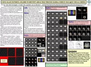
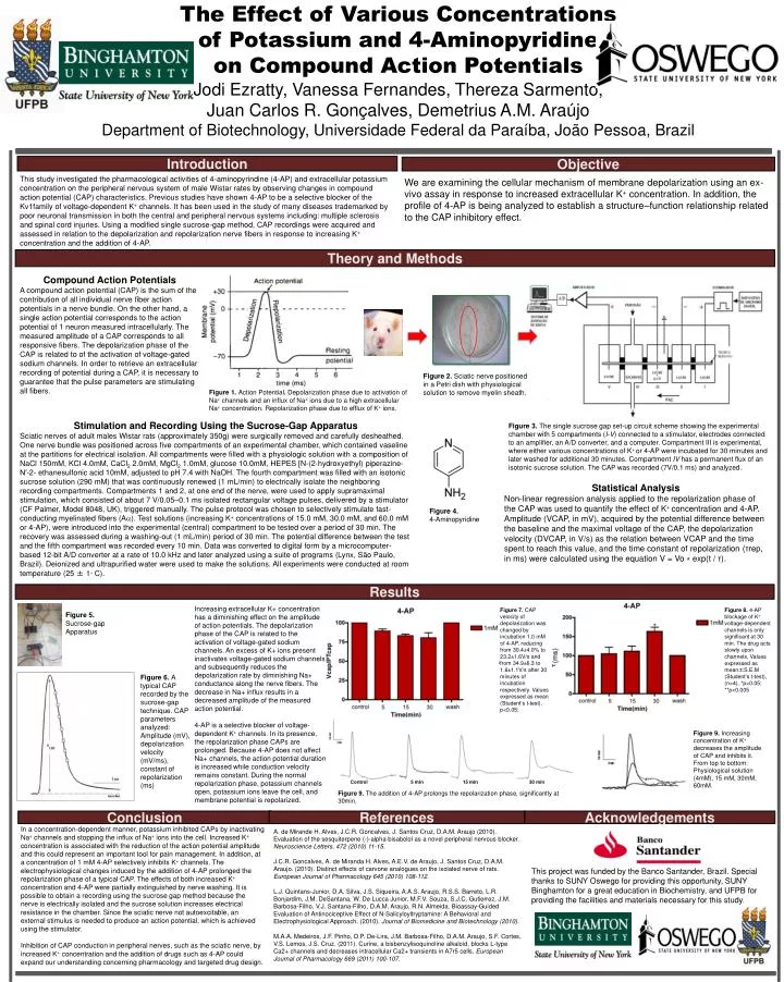
The Effect of Various Concentrations of Potassium and 4-Aminopyridine on Compound Action Potentials Jodi Ezratty, Vanessa Fernandes, TherezaSarmento, Juan Carlos R. Gonçalves, Demetrius A.M. Araújo Department of Biotechnology, Universidade Federal daParaíba, João Pessoa, Brazil Introduction Objective This study investigated the pharmacological activities of 4-aminopyrindine (4-AP) and extracellular potassium concentration on the peripheral nervous system of male Wistar rates by observing changes in compound action potential (CAP) characteristics. Previous studies have shown 4-AP to be a selective blocker of the Kv1family of voltage-dependent K+ channels. It has been used in the study of many diseases trademarked by poor neuronal transmission in both the central and peripheral nervous systems including: multiple sclerosis and spinal cord injuries. Using a modified single sucrose-gap method, CAP recordings were acquired and assessed in relation to the depolarization and repolarization nerve fibers in response to increasing K+ concentration and the addition of 4-AP. We are examining the cellular mechanism of membrane depolarization using an ex-vivo assay in response to increased extracellular K+ concentration. In addition, the profile of 4-AP is being analyzed to establish a structure–function relationship related to the CAP inhibitory effect. TheoryandMethods Compound Action Potentials A compound action potential (CAP) is the sum of the contribution of all individual nerve fiber action potentials in a nerve bundle. On the other hand, a single action potential corresponds to the action potential of 1 neuron measured intracellularly. The measured amplitude of a CAP corresponds to all responsive fibers. The depolarization phase of the CAP is related to of the activation of voltage-gated sodium channels. In order to retrieve an extracellular recording of potential during a CAP, it is necessary to guarantee that the pulse parameters are stimulating all fibers. Figure 2. Sciatic nerve positioned in a Petri dish with physiological solution to remove myelin sheath. Figure 1. Action Potential. Depolarization phase due to activation of Na+ channels and an influx of Na+ ions due to a high extracellular Na+ concentration. Repolarization phase due to efflux of K+ ions. Stimulation and Recording Using the Sucrose-Gap Apparatus Sciatic nerves of adult males Wistar rats (approximately 350g) were surgically removed and carefully desheathed. One nerve bundle was positioned across five compartments of an experimental chamber, which contained vaseline at the partitions for electrical isolation. All compartments were filled with a physiologic solution with a composition of NaCl 150mM, KCl 4.0mM, CaCl2 2.0mM, MgCl2 1.0mM, glucose 10.0mM, HEPES [N-(2-hydroxyethyl) piperazine-N′-2- ethanesulfonic acid 10mM, adjusted to pH 7.4 with NaOH. The fourth compartment was filled with an isotonic sucrose solution (290 mM) that was continuously renewed (1 mL/min) to electrically isolate the neighboring recording compartments. Compartments 1 and 2, at one end of the nerve, were used to apply supramaximal stimulation, which consisted of about 7 V/0.05–0.1 ms isolated rectangular voltage pulses, delivered by a stimulator (CF Palmer, Model 8048, UK), triggered manually. The pulse protocol was chosen to selectively stimulate fast-conducting myelinated fibers (A). Test solutions (increasing K+ concentrations of 15.0 mM, 30.0 mM, and 60.0 mM or 4-AP), were introduced into the experimental (central) compartment to be tested over a period of 30 min. The recovery was assessed during a washing-out (1 mL/min) period of 30 min. The potential difference between the test and the fifth compartment was recorded every 10 min. Data was converted to digital form by a microcomputer-based 12-bit A/D converter at a rate of 10.0 kHz and later analyzed using a suite of programs (Lynx, São Paulo, Brazil). Deionized and ultrapurified water were used to make the solutions. All experiments were conducted at room temperature (25 ± 1◦ C). Figure 3. The single sucrose gap set-up circuit scheme showing the experimental chamber with 5 compartments (I-V) connected to a stimulator, electrodes connected to an amplifier, an A/D converter, and a computer. Compartment IIIis experimental, where either various concentrations of K+ or 4-AP were incubated for 30 minutes and later washed for additional 30 minutes. Compartment IV has a permanent flux of an isotonic sucrose solution. The CAP was recorded (7V/0.1 ms) and analyzed. Statistical Analysis Non-linear regression analysis applied to the repolarization phase of the CAP was used to quantify the effect of K+ concentration and 4-AP. Amplitude (VCAP, in mV), acquired by the potential difference between the baseline and the maximal voltage of the CAP, the depolarization velocity (DVCAP, in V/s) as the relation between VCAP and the time spent to reach this value, and the time constant of repolarization (τrep, in ms) were calculated using the equation V = Vo ⁎ exp(t / τ). Figure 4. 4-Aminopyridine Results Increasing extracellular K+ concentration has a diminishing effect on the amplitude of action potentials. The depolarization phase of the CAP is related to the activation of voltage-gated sodium channels. An excess of K+ ions present inactivates voltage-gated sodium channels and subsequently reduces the depolarization rate by diminishing Na+ conductance along the nerve fibers. The decrease in Na+ influx results in a decreased amplitude of the measured action potential. 4-AP is a selective blocker of voltage-dependent K+ channels. In its presence, the repolarization phase CAPs are prolonged. Because 4-AP does not affect Na+ channels, the action potential duration is increased while conduction velocity remains constant. During the normal repolarization phase, potassium channels open, potassium ions leave the cell, and membrane potential is repolarized. Figure 7. CAP velocity of depolarization was changed by incubation 1.0 mM of 4-AP, reducing from 30.4±4.0% to 23.2±1.6V/s and from 34.9±6.3 to 1.6±1.1V/s after 30 minutes of incubation respectively. Valuesexpressed as mean (Student’st-test), p<0.05; Figure 8. 4-AP blockage of K+ voltage-dependent channels is only significant at 30 min. The drug acts slowly upon channels. Valuesexpressed as mean±S.E.M (Student’st-test), (n=4), *p<0.05; **p<0.005. Figure 5. Sucrose-gap Apparatus Figure 6. A typical CAP recordedbythesucrose-gaptechnique. CAP parametersanalyzed: Amplitude (mV), depolarizationvelocity (mV/ms), constantofrepolarization (ms) Figure 9. Increasing concentration of K+ decreases the amplitude of CAP and inhibits it. From top to bottom: Physiological solution (4mM), 15 mM, 30mM, 60mM. Control 5 min 15 min 30 min Figure 9. The addition of 4-AP prolongs the repolarization phase, significantly at 30min. Conclusion References Acknowledgements In a concentration-dependent manner, potassium inhibited CAPs by inactivating Na+ channels and stopping the influx of Na+ ions into the cell. Increased K+ concentration is associated with the reduction of the action potential amplitude and this could represent an important tool for pain management. In addition, at a concentration of 1 mM 4-AP selectively inhibits K+ channels. The electrophysiological changes induced by the addition of 4-AP prolonged the repolarization phase of a typical CAP. The effects of both increased K+ concentration and 4-AP were partially extinguished by nerve washing. It is possible to obtain a recording using the sucrose gap method because the nerve is electrically isolated and the sucrose solution increases electrical resistance in the chamber. Since the sciatic nerve not autoexcitable, an external stimulus is needed to produce an action potential, which is achieved using the stimulator. Inhibition of CAP conduction in peripheral nerves, such as the sciatic nerve, by increased K+ concentration and the addition of drugs such as 4-AP could expand our understanding concerning pharmacology and targeted drug design. A. de Mirande H. Alves, J.C.R. Goncalves, J. Santos Cruz, D.A.M. Araujo (2010). Evaluation of the sesquiterpene (-)-alpha-bisabolol as a novel peripheral nervous blocker. Neuroscience Letters, 472 (2010) 11-15. J.C.R. Goncalves, A. de Miranda H. Alves, A.E.V. de Araujo, J. Santos Cruz, D.A.M. Araujo. (2010). Distinct effects of carvone analogues on the isolated nerve of rats. European Journal of Pharmacology 645 (2010) 108-112. L.J. Quintans-Junior, D.A. Silva, J.S. Siqueira, A.A.S. Araujo, R.S.S. Barreto, L.R. Bonjardim, J.M. DeSantana, W. De Lucca Junior, M.F.V. Souza, S.J.C. Gutierrez, J.M. Barbosa-Filho, V.J. Santana-Filho, D.A.M. Araujo, R.N. Almeida. Bioassay-Guided Evaluation of Antinociceptive Effect of N-Salicyloyltryptamine: A Behavioral and Electrophysiological Approach. (2010). Journal of Biomedicine and Biotechnology (2010). M.A.A. Medeiros, J.F. Pinho, D.P. De-Lira, J.M. Barbosa-Filho, D.A.M. Araujo, S.F. Cortes, V.S. Lemos, J.S. Cruz. (2011). Curine, a bisbenzylisoquinoline alkaloid, blocks L-type Ca2+ channels and decreases intracellular Ca2+ transients in A7r5 cells. European Journal of Pharmacology 669 (2011) 100-107. This project was funded by the Banco Santander, Brazil. Special thanks to SUNY Oswego for providing this opportunity, SUNY Binghamton for a great education in Biochemistry, and UFPB for providing the facilities and materials necessary for this study.