Download
1 / 45
450 likes | 478 Views
Explore the process of cell division and its importance in development, limb regeneration, stem cell therapy, virology, and cancer. Learn how loss of cell division control leads to cancerous tumors and the role of oncogenes in cancer development.

E N D
Why Study Cell Division? • Development • Limb regeneration • Stem cells/stem cell therapy • Virology • Cancer
Cancer • Loss of cell division control leads to unlimited division --- cancerous tumors • Oncogenes are proteins that increase the chances of a normal cell becoming a cancer cell • Many viral proteins are oncogenes and links between virus infection and cancer have been found
Cancer http://www.nature.com/nrm/journal/v9/n8/box/nrm2457_BX1.html
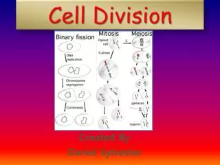
Cell Division • All cells arise from previously existing cells. • New cells are produced for growth or to replace damaged or old cells. • Cell division differs between prokaryotes and eukaryotes (binary fission vs. mitosis). • Cell division differs in asexual and sexual replication (mitosis vs meiosis).
Genetic Fidelity DNA contains all the information about cell function so each new cell must receive a complete, accurate copy of DNA during cell division. This ensures two identical daughter cells are produced.
Prokaryotic DNA In prokaryotes, a large circular chromosome is bound to the plasma membrane. Smaller plasmid DNA may also be present.
Chromosomes • Eukaryotic cells contain large amounts of genetic material. • The human cell contains more than 3 meters of DNA!!! • This DNA is packaged into structures called chromosomes located in the cell nucleus. • Most eukaryotes contain between 10 and 50 chromosomes. • Humans have 23 pairs (46) chromosomes.
Chromosome Structure DNA is wrapped around proteins called histones. http://cyberbridge.mcb.harvard.edu/dna_2.html http://www.mun.ca/biology/desmid/brian/BIOL2060/BIOL2060-18/CB18.html
Chromosome Structure • In dividing cells, chromosomes condense into tightly packaged chromatids. • In nondividing cells, chromosomes are more loosely packed and called chromatin.
Chromatids After DNA replication, the pair of sister chromatids are joined at the centromere. http://www.macroevolution.net/sister-chromatids.html http://www.uic.edu/classes/bios/bios100/lectures/mitosis.htm
Karyotype • Human chromosomes are paired and arranged by size in karyotypes. • The first 22 pairs are autosomes. • The final chromosome pair is the X/X or X/Y sex chromosomes. http://scigjt13.wordpress.com/2011/03/02/karyotype-of-alzheimers-disease/
Cell Reproduction • Asexual reproduction creates 2 diploid daughter cells that are identical to the parent cell. • Binary fission (prokaryotes); • Mitosis (eukaryotes) • Sexual reproduction creates daughter cells that are not genetically identical to the parent cell. • Meiosis
Prokaryotic Cell Reproduction Binary Fission
Binary Fission • Prokaryotes divide by binary fission. • Circular DNA chromosome (and plasmids) replicates. • Cell wall forms creating two new, smaller cells. • Animation http://academic.pgcc.edu/~kroberts/Lecture/Chapter%206/fission.html
Eukaryotic Cell Cycle • Two Stages of Eukaryotic Cell Cycle • Interphase • G1 – Primary growth phase • S phase – DNA synthesis • G2 – Secondary growth phase • Mitosis and Cytokinesis
The Cell Cycle http://www.bristol.k12.ct.us/page.cfm?p=7094
Interphase: The G1 Stage • 1st stage of growth for a new cell after division • Cells mature (make more cytoplasm and organelles). • Cells will carry out normal metabolic and biological functions.
Interphase: The S Phase • S for DNA Synthesis • DNA is replicated to create 2 complete copies of original chromosomes
Interphase: The G2 Stage • 2nd stage of growth following DNA replication • Cell creates all organelles and proteins that will be needed for cell division • Example - centrioles
Interphase: Summary • Newly formed cells grow in size and make organelles/proteins needed to perform function (G1). • Chromosomes replicate making 2 complete copies (S). • Cells make organelles and proteins needed for mitosis (G2).
Mitosis • Division of nucleus • Occurs in eukaryotes only • Consists of 4 steps: • Prophase • Metaphase • Anaphase • Telophase • Does not occur in some cell types (nerve cells) http://botit.botany.wisc.edu/Resources/Botany/Mitosis/Allium/Complete%20Mitosis.jpg.html
Mitosis – Early Prophase • Chromatin condenses to form visible chromosomes • Mitotic spindle forms from fibers in cytoskeleton or centrioles cell wall nucleolus chromatin nuclear envelope http://www.bristol.k12.ct.us/page.cfm?p=7094 http://mrteacherdude.com/biology/biology2/lab_stuff/mitosis/mitosis_plant_pro.htm
Mitosis – Late Prophase • Nuclear membrane and nucleolus are broken down. • Chromatin continues to condense. • Kinetochores attach to centromeres. • Spindle forms at pole of cell. http://www.bristol.k12.ct.us/page.cfm?p=7094 http://mrteacherdude.com/biology/biology2/lab_stuff/mitosis/mitosis_plant_pro.htm
Kinetochore and Spindle Fibers • The mitotic spindle forms from microtubules in plants and centrioles in animals. • Polar fibers extend from one pole of the cell to the other. • Short asters radiate from spindle. http://en.wikipedia.org/wiki/Spindle_apparatus http://en.wikipedia.org/wiki/Spindle_apparatus
Kinetochore and Spindle Fibers http://en.wikipedia.org/wiki/Spindle_apparatus Kinetochore fibers extend from the pole to the centromere of the chromosome.
asters pole chromosomes
Mitosis - Metaphase • Chromosomes attached to kinetochores move to the center of the cell. • Lined up at equator. equator pole http://www.bristol.k12.ct.us/page.cfm?p=7094 http://www.cbv.ns.ca/bec/science/cell/page17.html
Mitosis - Anaphase • Kinetochore fibers pull sister chromatids apart. • Very rapid. http://www.bristol.k12.ct.us/page.cfm?p=7094 http://micro.magnet.fsu.edu/micro/gallery/mitosis/earlyanaphase.html
Mitosis - Telophase • Sister chromatids migrate to opposite poles. • Spindle disassembles. • Nuclear envelopes form around each set of sister chromatids. • Nucleolus reappears. • Chromosomes return to chromatin. • Cytokinesis occurs. http://www.bristol.k12.ct.us/page.cfm?p=7094 http://botit.botany.wisc.edu/Resources/Botany/Mitosis/Allium/telophase%20cytokinesis.jpg.html
Mitosis - Cytokinesis • Division of cytoplasm between daughter cells • In plants a cell plate forms at the equator to divide cells. • In animals a cleavage furrow divides cells.
Mitosis - Cytokinesis Cleavage Furrow Cell Plate http://www.uic.edu/classes/bios/bios100/lectures/mitosis.htm http://kr021.k12.sd.us/mitosis_practice_test.htm http://www.vcbio.science.ru.nl/en/virtuallessons/mitostage/
Completion of Mitosis • After mitosis you have 2 daughter cells that are identical to parent cell in chromosome number and DNA sequence*. • Daughter cells are smaller than mature cell and enter G1 phase (interphase) to grow and function. http://faculty.clintoncc.suny.edu/faculty/michael.gregory/files/bio%20101/bio%20101%20lectures/mitosis/mitosis.htm
Mitosis Mitosis Animation Plant Cell Mitosis
Mitosis Quiz 1. The 2 chromatid arms are held together in the center by a _____________. A. centrosome B. centriole C. centromere D. histone
Mitosis Quiz 2. A cell with only one of each kind of chromosome is said to be 1n or ______. A. diploid B. cancer C. haploid D. mitotic
Mitosis Quiz 3. Which of the following shows the correct order for the phases of the cell cycle? A. G1 – G2 – S – M B. S – G1 – M – G2 C. G1 – S – G2 – M D. G1 – M – G2 - S
Mitosis Quiz 4. The diagram shown is a picture of a person’s chromosomes called a _______. A. karyotype B. genome C. chromatid D. centromere 5. The person shown is _____ A. Male B. Female
Mitosis Quiz 6. Cells spend most of their time in this stage of the life cycle. A. Interphase B. Mitosis C. Telophase D. Anaphase
Mitosis Quiz 7. The place where the cell membrane pinches in to make 2 new daughter cells is called the _______. A. cleavage furrow B. cell plate C. pole D. kinetochore
A. B. C. D. Mitosis Quiz Which of the pictures shows: 8. Metaphase? 9. Telophase? 10. Prophase? 11. Anaphase