Download
1 / 17
170 likes | 313 Views
膝关节磁共振检查. 西南医院关节外科中心. 介绍 MRI 之前需要注意的问题. X 线成像( X-ray )是膝关节的影象学基础,好的 X 线片可以提供大量的重要信息。 不仅是骨性结构,软组织病变也可以反映在在 X 线片上. CR 机( Computed Radiography )的出现将 X 线片在疾病诊断中的应用进一步扩大. CT ( Computed Tomograghy )对大部分膝关节病变的显示没有比 X 线片更明显的优越性. 在三维空间结构的识别上有可借鉴之处. MRI ( Maganetic resonance imaging )是相对容易掌握的影象学检查

E N D
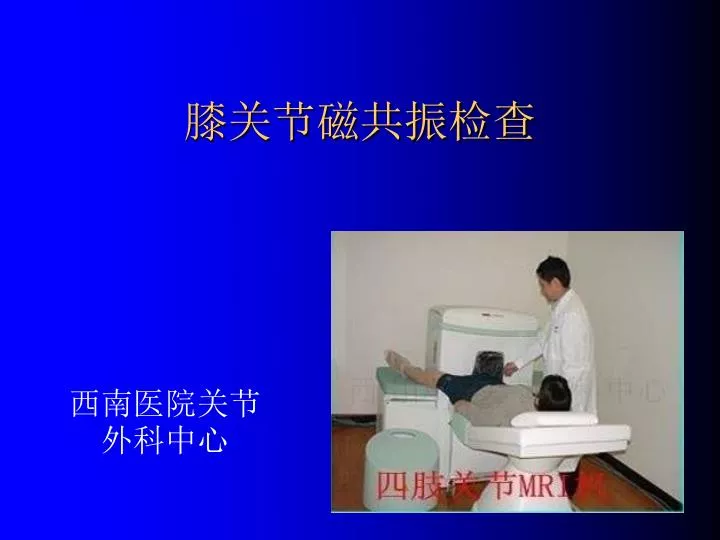
膝关节磁共振检查 西南医院关节外科中心
介绍MRI之前需要注意的问题 • X线成像(X-ray)是膝关节的影象学基础,好的X线片可以提供大量的重要信息。 • 不仅是骨性结构,软组织病变也可以反映在在X线片上
CR机(Computed Radiography)的出现将X线片在疾病诊断中的应用进一步扩大
CT(Computed Tomograghy)对大部分膝关节病变的显示没有比X线片更明显的优越性
MRI(Maganetic resonance imaging)是相对容易掌握的影象学检查 • 对软组织的显示最具有优势
MRI 原理 • 氢原子含有一个质子,容易受外加磁场作用产生磁共振现象 • 人体不同组织含有的氢原子数量不同 • 磁场---激励状态---弛豫状态---释放电磁波---接受成像 • 简言之:MRI是利用氢原子在磁场中被激励后释放能量的被接受形成的图象。
T1:显示解剖结构好,图象细腻 • T2:显示病变结构好,噪声比例高