Download
1 / 17
440 likes | 1.21k Views
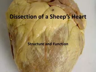
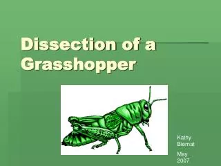
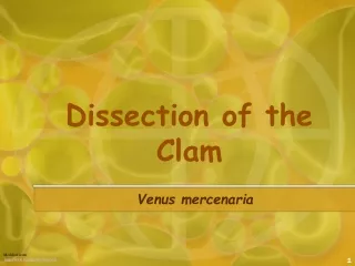
Dissection of a Clam. Phylum Mollusca, Class Bivalvia. Photos by Dr. J. Whaley Flagler Palm Coast High School 2013. A variety of clams are used for dissection. Therefore, students should expect to see some variation in shell shape and internal structures. Dorsal Aspect- along the back.

E N D
Dissection of a Clam Phylum Mollusca, Class Bivalvia Photos by Dr. J. Whaley Flagler Palm Coast High School 2013
A variety of clams are used for dissection. Therefore, students should expect to see some variation in shell shape and internal structures. Dorsal Aspect- along the back Umbo Hinge Ligament Posterior View- toward the rear or anus (area of the siphons) Anterior View- toward the head Ventral Aspect- toward the belly
After the adductor muscles have been cut, the valves of the clam can be opened slowly. This allows the mantle to be viewed as it lines the the shell. The mantle contains the shell glands which produce the shell and it protects the internal organs. The left and right mantle form the siphons. They are contracted in the shell in a preserved specimen. There is a picture of a live clam from the class tank which shows the siphons extended. When disturbed the clam will withdraw them into shell. Posterior View- see the mantle forms the siphons. Ventral View- note the mantle coming together on the posterior aspect to form the siphons. The Adductor Muscles and the Foot are visible.
Live Clam Siphons
Ventral Views The posterior aspect is on the right side of each picture.
Mantle Cavity – with the visceral mass (shell removed) Note: how the mantle is thin in a portion of the structure and thick along the edge where the shell glands are located. Anterior Aspect Posterior Aspect
Internal Anatomy of a Clam Palps Adductor Muscle Visceral Mass Foot Mantle Hinge Ligament Gills Adductor Muscle Siphon Region
Different Clam Species – a different look.. Adductor Muscle Palps Foot Mantle Gills Siphon Region Visceral Mass
Siphon Region Excurrent Siphon Incurrent Siphon
Gills have been lifted up to expose the visceral mass Gills- left side Palps Foot Gills- right side Visceral Mass
Close-up of the gills. The gills are used for both respiration and feeding.
To access the visceral mass… cut off the muscular foot, then cut down through the visceral mass to create a left and right side
Visceral Mass has been cut open. Green area is the digestive glands that surround the stomach. The cream colored area are the reproductive structures which surround the intestines. Intestines Gonads Digestive Glands and stomach region
Different Species –different look Adductor Muscle
Anterior View The palps collect food from the gills and then form a mass to direct the food into the mouth in the center. Right Palp Foot Left Palp
Dorsal View Anterior Left Side Right Side Heart Region The intestine runs through this area before emptying at the anus. Kidney Region Posterior