Download
1 / 10
110 likes | 281 Views
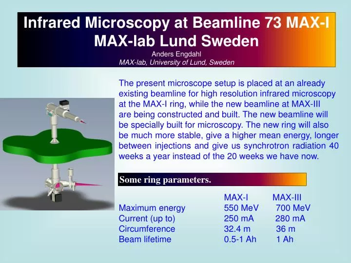
Infrared Microscopy at Beamline 73 MAX-I MAX-lab Lund Sweden Anders Engdahl MAX-lab, University of Lund, Sweden. The present microscope setup is placed at an already existing beamline for high resolution infrared microscopy at the MAX-I ring, while the new beamline at MAX-III

E N D
Infrared Microscopy at Beamline 73 MAX-I MAX-lab Lund Sweden Anders Engdahl MAX-lab, University of Lund, Sweden The present microscope setup is placed at an already existing beamline for high resolution infrared microscopy at the MAX-I ring, while the new beamline at MAX-III are being constructed and built. The new beamline will be specially built for microscopy. The new ring will also be much more stable, give a higher mean energy, longer between injections and give us synchrotron radiation 40 weeks a year instead of the 20 weeks we have now. Some ring parameters. MAX-I MAX-III Maximum energy 550 MeV 700 MeV Current (up to) 250 mA 280 mA Circumference 32.4 m 36 m Beam lifetime 0.5-1 Ah 1 Ah
Beamline Equipment Spectrometer IFS/66V vacuum instrument: Beamsplitters: Near Infrared: Si/CaF2. Mid Infrared : Ge/KBr, Far Infrared: 6 μ mylar Sources: NIR: Tungsten lamp, Mid Infrared: Globar All spectral regions: Synchrotron radiation. Hyperion 3000 microscope. Detectors: InSb: NIR 10000-1850 cm-1.MCT: mid IR with detector area 0.25*0.25 mm 10000-600 cm-1. MCT: mid IR with detector area 0.5*0.5 mm 10000-600 cm-1. Si-Bolometer: Far IR, 600 -10 cm-1. FPA mid IR (Photovoltaic MCT Focal Plane Array Detector). 128*128 elements. Objectives: Grazing angle 20x IR, ATR Germanium IR, 100 micron contact area. ATR Silica IR including FIR, 300 micron contact area. 36x IR, 15x IR, 4x glass visible Transmittance and reflection modes possible for the 36x, 15x and 4x objectives
Identification of organic tissue in fossile bones A mosasaur (a sea living animal, the species we have looked at is around 70 000 000 years old) The sample that triggered this investigation. The long structures are blood vessels so the discovery of blood-like matter inside vessels Arouse an interest in what it might be. The upper forearm bone that we are now looking at.
Optical microscope picture Infrared microscope picture (Integration over the CH-stretching region.) A few example spectra from the system.
Projects Localization of enzymes in granulates A study of the localization of enzymes in a variety of solid products. An understanding of the distribution of the enzyme within the particles may make it possible to understand and improve production processes. It can also give insight ín why enzymes are more stable in certain particles than in other. By using the characteristic infrared bands of the amide bonds of proteins, enzymes such as Lipozyme TL IM can be localized in thin cross sections of particles. This project is on hold for now but will be continued later. To the left: Optical picture of a particle. To the right: Infrared spectroscopic picture With integration from 1489 to 1572 cm-1 showing the concentration of the enzyme.
Identification of particles in ointment Sometimes it is a complicated task to extract and identify particles in an ointment. With the FPA-detector it is possible to solve problems like this. In the present case the ointment cotained two active substances. The ointment is applied on an ir-transparent material in a thin layer, and then the sample is put in the microscope to be examined Integration over a band from the other substance. Optical picture Integration over an absorption band from one of the substances in the ointment.
Indoor corrosion on copper Indoor corrosion is a problem in for example the electronics industy and in the preservation of old copper art objects. Copper plates treated in a known atmosphere of humid air and a known concentration of either acetic acid or formic acid for different periods of time were analysed both optically and spectroscopically to get a knowledge of the local variations of corrosion. Left picture: Corroded copper: Middle picture: Integration1521 to 1623 cm-1. Right picture: Integration: 1295 to 1355 cm-1.
Biological aspects of water structure in human and animal skin Far-IR spectra contain information about intermolecular interactions in hydrogen bonded systems of biological interest. Vibrations arising from hydrogen bonded intermolecular interactions are found at low frequencies due to the weak intermolecular forces and heavy masses involved in the vibrations. Vibrational excitations of low wavenumber collective modes are essential for important conformational transitions in proteins. In this context a broad band around 115 cm-1 is important. We assigned this band to an out-of-plane mode of atoms in the peptide hydrogen bonds. These modes may collectively drive a conformational transition of a protein. The figure shows Far-IR and low wavenumber Raman spectra of polyglycine. We have quite recently been able to see the influence On some protein bands upon adding of water. Also Pure water absorptions were observed. Synchrotron radiation is a very powerful light source in the Far-IR region. In another project a characterization of far infrared vibrations for different biomolecules have been started.
Chemical imaging of a mouse brain A mouse brain sliced in 10 my thick horisontal slices has been examined by infrared microscopy. A number of reference substances has been measured, and by integration over regions where the individual substances has absorptions an infrared image of the brain has been obtained. Cholesterol is among the investigated substances and it is evident that we can see the distribution of cholesterol in the brain tissue by this method. The two pictures to the left show integration over two different cholesterol absorbtions. Picture three shows a region where another substance has an absorbtion band and the picture to right a region where no absorptions at all are found.
Some recently started projects. Speciation in preserved water-logged archeological wood from the Vasa. Some preliminary tests have been done.. A model system for studies of diffusion of nutrients in bone tissues using infrared microspectroscopy. Tests to see model substances in bone has been performed. Chemical mapping of single aerosol particles in an electrodynamic balance particle trap. A balance has been constructed. Has only been tested with a model substance with very low vapor pressure so far.