Download

1 / 56
670 likes | 1.19k Views
THE PATHOLOGY OF THE ADRENAL GLAND. ANATOMY. The adrenal glands are endocrine organs consisting of both cortex and medulla. ANATOMY. Adrenal cortex: the narrow layer of zona glomerulosa ( mineralocorticoids)

E N D
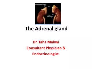
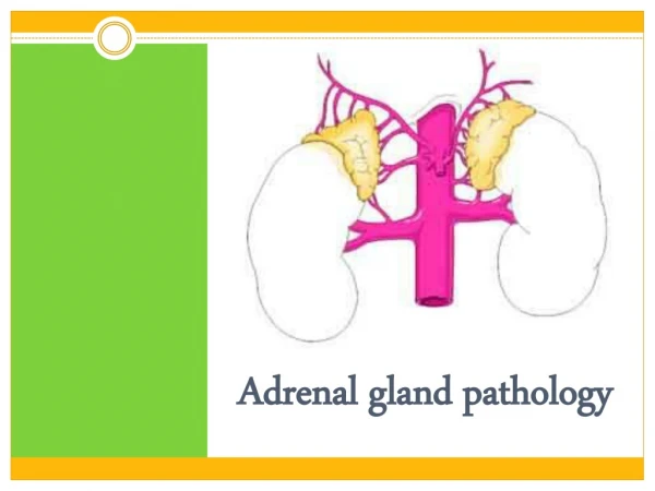
ANATOMY The adrenal glandsare endocrine organs consisting of both cortex and medulla.
ANATOMY Adrenal cortex: the narrow layer of zona glomerulosa ( mineralocorticoids) Intervening is the broad zona fasciculata, which makes up about 75% of the total cortex ( glucocorticoids) The narrow layer of zona reticularis abuts the medulla (sex steroids)
ANATOMY The adrenal medulla is composed of chromaffin cells, which synthesize and secrete catecholamines, mainly epinephrine.
ANATOMY The adrenal cortex synthesizes three different types of steroids: (1) glucocorticoids (principally cortisol), which are synthesized primarily in the zona fasciculata and to a lesser degree in the zona reticularis; (2) mineralocorticoids, the most important being aldosterone, which is generated in the zona glomerulosa; (3) sex steroids (estrogens and androgens), which are produced largely in the zona reticularis
Developmental anomalies Adrenal Hypoplasia: Anencephalic type: may have adrenal agenesis. Cytomegalic type: Large eosinophilic cells, may be X-linked. Ectopic Adrenal: Accessory adrenal (cortex) can be found from diaphragm to pelvis in the retroperitoneum (Medulla can also be found if near celiac ganglion). May be seen in hernial sacs (Marchand rests). These may give origin to neoplasms or undergo hyperplasia. Adrenal myelolipoma: a metaplasia-choristoma made of bone marrow. They can be big but are generally harmless.
ADRENOCORTICAL HYPERFUNCTION (HYPERADRENALISM) There are three basic types of hyperadrenal syndromes: (1) Cushing syndrome, characterized by an excess of cortisol; (2) hyperaldosteronism; (3) adrenogenital or virilizing syndromes caused by an excess of androgens.
Hypercortisolism (Cushing Syndrome) Cushing syndrome can be broadly divided into exogenousand endogenouscauses. The vast majority of cases of Cushing syndrome are the result of the administration of exogenous glucocorticoids ("iatrogenic" Cushing syndrome).
Hypercortisolism (Cushing Syndrome) The endogenous causes can, in turn, be divided into those that are ACTH dependent and those that are ACTH independent
Hypercortisolism (Cushing Syndrome) ACTH-DEPENDENT Cushing disease (pituitary adenoma; rarely CRH-dependent pituitary hyperplasia) Ectopic corticotropin syndrome (ACTH-secreting pulmonary small-cell carcinoma, bronchial carcinoid) ACTH-INDEPENDENT Adrenal adenoma Adrenal carcinoma Macronodular hyperplasia (ectopic expression of hormone receptors, including GIPR, LHR, vasopressin and serotonin receptors) Primary pigmented nodular adrenal disease (PRKARIA and PDE11 mutations) McCune-Albright syndrome (GNAS mutations)
Cushing's Syndrome Middle-aged 3F/1M 4 main causes: Pituitary adenoma - secretes ACTH Adrenal tumors - secrete cortisol Ectopic production of ACTH Iatrogenic Cushing's Syndrome
Pituitary adenoma - secretes ACTH- 60-70% of cases of pathologic causes- adrenal glands are hyperplastic- Biochemistry: Increased ACTH and cortisol
2. Adrenal tumors secrete cortisol 20-25% of cases of Cushing's adrenal adenoma (50%), carcinoma (50%) atrophy of contralateral gland Biochemistry: low ACTH and high cortisol
3. Ectopic production of ACTH by tumors lung cancer, bronchial and thymic carcinoids, medullary thyroid carcinoma, islet cell cancer 10-15% of cases adrenals are hyperplastic Biochemistry: Increased ACTH and cortisol
4. Iatrogenic Cushing's Syndrome commonest cause overall due to long-term corticosteroid therapy for treatment of:- connective tissue diseases - asthma- rheumatoid arthritis - cancer- transplant rejection- Biochemistry: High cortisol and ACTH suppressed
Clinical findings in Cushing's Syndrome Occurs as a consequence of excess plasma glucocorticoid levels truncal obesity, buffalo hump and moon face due to fat deposition hypertension hirsutism - due to increased testosterone muscle weakness and breakdown menstrual irregularity osteoporosis - decreased calcium absorption, increased bone resorption and suppression of bone formation impaired glucose tolerance or diabetes mellitus - due to insulin resistance abdominal striae - due to inhibition of protein synthesis and abnormal collagen maturation mental symptoms are common; depression and psychosis
Primary adrenal hyperplasia a newly discovered disease: "primary pigmented nodular adrenocortical hyperplasia", which is part of the autosomal dominant Carney complex (Carney complex: cardiac myxomas, cutaneous myxomas, spotty pigmentation of the skin, and hyperendocrine states) genetic syndrome with too many cortisol receptors, low plasma cortisol macronodular hyperplasia with marked adrenal enlargement
Primary Hyperaldosteronism Hyperaldosteronismis the generic term for a group of closely related conditions characterized by chronic excess aldosterone secretion.
Primary Hyperaldosteronism Hyperaldosteronism may be primary, or it may be secondary to an extra-adrenal cause. Primary hyperaldosteronismstems from an autonomous overproduction of aldosterone, with resultant suppression of the renin-angiotensin system and decreased plasma renin activity. Blood pressure elevation is the most common manifestation of primary hyperaldosteronism, which is caused by one of three mechanisms:
Primary Hyperaldosteronism Bilateral idiopathic hyperaldosteronism (IHA), Adrenocortical neoplasm, Glucocorticoid-remediable hyperaldosteronism
Bilateral idiopathic hyperaldosteronism (IHA) Bilateral idiopathic hyperaldosteronism (IHA), characterized by bilateral nodular hyperplasia of the adrenal glands, is the most common underlying cause of primary hyperaldosteronism, accounting for about 60% of cases. Individuals with IHA tend to be older and to have less severe hypertension than those presenting with adrenal neoplasms.
Adrenocortical neoplasm Either an aldosterone-producing adenoma (the most common cause) or, rarely, an adrenocortical carcinoma. In approximately 35% of cases, primary hyperaldosteronism is caused by a solitary aldosterone-secreting adenoma, a condition referred to as Conn syndrome. This syndrome occurs most frequently in adult middle life and is more common in women than in men (2 : 1). Multiple adenomas may be present in an occasional patient.
Glucocorticoid-remediable hyperaldosteronism Glucocorticoid-remediable hyperaldosteronism is an uncommon cause of primary familial hyperaldosteronism. In some families, it is caused by a chimeric gene resulting from fusion between CYP11B1 (the 11β-hydroxylase gene) and CYP11B2 (the aldosterone synthase gene). This leads to a sustained production of hybrid steroids in addition to both cortisol and aldosterone. The activation of aldosterone secretion is under the influence of ACTH and hence is suppressible by exogenous administration of dexamethasone.
Primary hyperaldosteronism low-renin hyperaldosteronism too much mineralocorticoid not due to excess ACTH 0.5% of hypertensives have primary hyperaldosteronism Patients exhibit hypokalemia, alkalosis, and low renin, Low potassium is likely to cause muscle weakness, and even paralysis. these patients can die from hypokalemia if you give them thiazide diuretics to treat their high blood pressure.
Secondary aldosteronism In secondary hyperaldosteronism, in contrast, aldosterone release occurs in response to activation of the renin-angiotensin system. It is characterized by increased levels of plasma renin and is encountered in conditions such as the following: Decreased renal perfusion (arteriolar nephrosclerosis, renal artery stenosis) Arterial hypovolemia and edema (congestive heart failure, cirrhosis, nephrotic syndrome) Pregnancy (due to estrogen-induced increases in plasma renin substrate)
Hypofunction of adrenal cortex Primary causes Loss of Cortex: autoimmune bacterial hemorrhage amyloidosis sarcoidosis hemochromatosis metastatic disease surgical ablation. Metabolic Failure: Primary failure of adrenal glands, Drug induced inhibition of adrenocortical function.
Secondary causes hypothalamic / pituitary disease neoplasm infection hypothalamic / pituitary suppression: long term steroid administration.
AdrenocorticalInsufficiency(Hypoadrenocorticism) Primary Acute Adrenal Insufficiency: Precipitated by stress in a patient with Addison's. Precipitated by withdrawal of steroids in an adrenalectomized patient. In patients with acute bilateral hemorrhagic necrosis of adrenals. Chronic insufficiency: Addison's disease
Acute Insufficiency Acute Hemorrhagic Necrosis of Adrenals Waterhouse-Friedricksen Syndrome Due particularly to meningococcal septicemia, less commonly Staph, pneumococcus, or Haemophilus Newborns due to perinatal trauma. DIC adrenal glands are hemorrhagic with cortical necrosis purpuric rash vascular collapse shock: due to endotoxin and vomiting, decreased steroids and Na+, increased K+ in blood and dehydration.
Primary chronic adrenal insufficiency Addison's disease 60-75% due to auto-immune destruction of adrenal cortex with lymphocytic and plasma cell infiltrate normal medulla Tuberculosis (2nd commonest cause) more than 90% of cortex needs to be destroyed ClinicallyLethargy, muscle weakness, anorexia, nausea & vomiting, wt loss, hypotension, hyperpigmentation of skin and mucous membranes.
Primary: due to adrenal insufficiency should be suspected when there is marked skin pigmentation due to high ACTH levels. Secondary: pituitary or hypothalamic insufficiency (no skin pigmentation). Addison's disease is rare.
Causes of Addison's Disease 1. Autoimmune most cases of Additions show anti-adrenal Antibodies (60-75%) Two types exist. Type I -Addison's + hypoparathyroidism+ mucocutaneous candidiasis. -Defect in suppressor cells.
Type II(Schmidt's syndrome) Addison's + autoimmune thyroid disease + insulin dependent diabetes. No Candidiasis or PTH deficiency Associated with HLA-A1 and HLA-B8 haplotype. 2. Tuberculosis (~25% of cases). 3. Other causes: histoplasmosis amyloid sarcoidosis metastases • congenital hypoplasia • hemochromatosis • CMV infection (in AIDS) • adrenal leukodystrophy
Pathology There may be a lymphocytic infiltrate or a "burned out" fibrotic appearance. Medulla is untouched. In TB and other diseases, evidence for these is usually apparent.
Adrenal Medulla Pathology Adrenal Medulla Synthesizes and secretes "adrenalin" (epinephrine, norepinephrine) The only diseases are two tumors which may arise here or at the other chromaffin tissue masses pheochromocytoma (well-differentiated, adults) neuroblastoma (poorly-differentiated, children).
Pheochromocytoma(paraganglioma) secrete norepinephrine (most common) and/or epinephrine Rarely: dopamine, serotonin, ACTH, somatostatin, neuropeptides. The paroxysms of extreme hypertension, accompanied by sweating, headache, and other autonomic disturbances, probably result from physical compression and/or ischemia of the "pheo".
Can present as Paroxysmal attacks which may precipitate congestive cardiac failure, coronary thrombosis, cerebral hemorrhage may be brought on by stress, pressure, exercise acute release of catecholamine Biochemistry: increased urinary VMA (vanillylmandelic acid) excretion
Most (90%) occur in the adrenal medulla Other sites (10%) the organs of Zuckerkandl: chromaffin tissue at the origin of the inferior mesenteric artery and/or aortic bifurcation paravertebral sympathetic chain urinary bladder.
Ten percent of pheo cases involve both adrenals, and many of these are also among the 10% that are familial. Syndromes include: MEN IIa or IIb von Recklinghausen's neurofibromatosis (NF-I) von Hippel-Lindau succinic dehydrogenase mutation
Pheos are still often missed clinically surprises at autopsy the patients typically had been told they had "benign essential hypertension" and "emotional problems“ high levels of circulating catecholamines can damage the myocardium can cause coronary spasm, disorders in renal, bowel, brain arteries
Grossly, very bloody (vascular), often show fibrosis, calcification, cystic change, or even fatty change Microscopically, pheos resemble adrenal medulla special stains are available that show norepinephrine and/or epinephrine in granules
Neuroblastoma The second commonest solid pediatric cancer (after Wilms tumor). It is derived from primitive nerve elements, discovered by Virchow. A majority of neuroblastomas arise in or near the adrenals. Grossly, neuroblastomas are soft, white tumors.