Download
1 / 18
180 likes | 350 Views
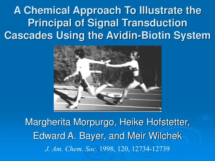
A Chemical Approach To Illustrate the Principal of Signal Transduction Cascades Using the Avidin-Biotin System. Margherita Morpurgo, Heike Hofstetter, Edward A. Bayer, and Meir Wilchek. J. Am. Chem. Soc. 1998, 120, 12734-12739. Introduction.

E N D
A Chemical Approach To Illustrate the Principal of Signal Transduction Cascades Using the Avidin-Biotin System Margherita Morpurgo, Heike Hofstetter, Edward A. Bayer, and Meir Wilchek J. Am. Chem. Soc. 1998, 120, 12734-12739
Introduction In nature, cellular functions are propagated by cascades of molecules, which interact with one another for signal transduction. Generally, the sequential process is initiated by the binding of an extracellular signal to a receptor culminating in one or more specific cellular responses In this way, a signal, for example, can be transferred from the outside of the cell en route to the nucleus—a process mediated by ligands, e.g., hormones, cytokines, and growth factors.
Goal To determine whether a cascade can be formed artificially, whereby the binding of one molecule would depend on the “signaling” of another. Two molecules that display differing affinities for the same binding site of a protein: [Simple diagram] Consequently, one step will be dependent on a previous one, thus enabling us to trigger a cascade of binding, e.g., to construct an organized system of protein multilayers.
Background Method based on the Avidin-Biotin system: Avidin-biotin is based on the high affinity of the cofactor Biotin for the protein Avidin and the ease of spectrophotometric measurement Used the egg-white protein avidin, deglycosylated (DG) avidin, and streptavidin which bind to two different ligands, biotin and 4-hydroxyazobenzene-2-carboxylic acid (HABA), with different affinities, 10-15 and 10-6 M, respectively.
Background Based on the binding of the dye HABA to Avidin and the ability of Biotin to displace the dye in stoichiometric proportions Binding of HABA to avidin is accompanied by an instantaneous shift in the absorption spectrum from Ïmax 348 nm of the free HABA to Ïmax 500 nm of the complexed tautomer. Furthermore, avidin can be forced to catalyze the hydrolysis of HABA derivatives, in which the hydroxyl function is blocked with a protecting group (e.g., an acetyl moiety), to accommodate the azo dye in its tautomeric form at the binding site.
HABAylated avidin + antibody + biotin Biotin (blue) binds avidin and expells HABA Anti-HABA antibodies + antibody + avidin B Biotinylated antibody The Plan, Stan:
Cyclic HABA OH- Hydrolysis A HABA-containing affinity label was designed, such that the dye would remain covalently attached to the binding site of avidin.
w/biotin w/o biotin Cyclic HABA added to avidin. Reaction monitored by UV-Vis spec. Gradual shift in absorption from 328 nm to 504 nm
Biotin was added to the red HABAylated avidin: immediate shift to 356 nm observed Yet, the orange color (of HABA) continued to be associated with the protein, indicating HABA was still covalently bound to colorless avidin.
More evidence of HABA covalently binding avidin Avidin + HABA m/z = 14 727 Difference: 14 727 -14 289 438 m/z = 14 289 ~single subunit of avidin ~ mass of cyclic HABA Matrix-Assisted Light Desorption Ionization (MALDI) Mass Spectra of avidin before and after reaction with cyclic HABA.
Amino Acid Sequence of HABAylated Avidin (orange) HPLC analysis of the tryptic digest of HABAylated avidin. To determine which avidin residue was modified and covalently bound to the HABA moiety, the protein was subjected to trypsin digestion. 356 nm, adsorption maxima of HABA 220 nm, adsorption maxima of the peptides. Lys-111
Cyclic HABA can be hydrolyzed by avidin, which enables the attachment of the HABA moiety to an appropriate residue in or near the binding site of avidin. HABA with side chain Adjacent monomer on avidin Binding site of avidin (one monomer)
Polyclonal antibodies against HABA were examined for their interaction with the HABAylated avidin in the presence and absence of biotin. w/ biotin w/o biotin
MissionComplete HABAylated avidin + biotin B Biotin (blue) binds avidin and expells HABA Biotin-saturated HABAylated Avidin + Biotinylated antibody B
In Summary… • Used the avidin-biotin system to demonstrate the interplay of molecular recognition and oriented protein assembly which serves as a prerequisite for the principle of signal transduction • Biotin served as the effector or trigger of the cascade • Avidin has four binding sites: limited amounts of biotin can be added to displace only one or two HABA molecules to trigger the cascade vectorially in different dimensions
Conclusion • System can be considered a chemical mimic of signal transduction, which, in nature, can also be modulated in different ways to enhance the response, following the initial triggering by a hormone or another effector • This concept of chemical mimicry and the assembly of protein multilayers is appropriate for application in numerous fields, such as medicine, diagnostics, biosensors, nanotechnology, and artificial intelligence, thus expanding the scope of the avidinbiotin system.