Download

1 / 44
460 likes | 481 Views
27.4.2015. SKIN AND ITS APPENDAGES. Dr. Archana Rani Associate Professor Department of Anatomy KGMU UP, Lucknow. INTRODUCTION. Skin is the outer covering of the body. Skin and its appendages constitute the integumentary system . Largest organ of the body.

E N D
27.4.2015 SKINAND ITS APPENDAGES Dr. Archana Rani Associate Professor Department of Anatomy KGMU UP, Lucknow
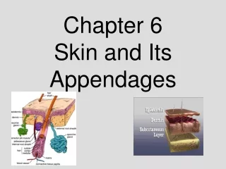
INTRODUCTION • Skin is the outer covering of the body. • Skin and its appendages constitute the integumentary system. • Largest organ of the body. • Constitutes 16% of the body weight.
Some Facts about Skin • Surface area: 1.5-2.0 sq meters • Thickness: 0.5-3.0 mm • Growth rate of nail: 0.1mm per day • Growth rate of hair: 1.5-2.2 mm per week • Life span of hair: Eyelashes, axilla- 4 months Scalp – 4 years
FUNCTIONS OF SKIN • Protective shield for the body • Barrier to water • Thermoregulation • Important sense organ (pain, touch, temperature & pressure) • Absorption of ultraviolet radiation from sun for the production of vitamin D
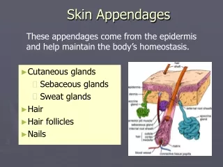
LAYERS OF THE SKIN Epidermis: • Composed of keratinized stratified squamousepithelium. Dermis: • Papillary region composed of loose connective tissue. • Reticular region composed of dense irregular connective tissue. Hypodermis: • Composed of areolar tissue with abundant adipocytes.
LAYERS OF THE EPIDERMIS Stratum Basale (Germinal/ Malpighian layer): • Single layer of cuboidal cells resting on basement membrane. • High mitotic activity. Stratum Spinosum: • Several layers of polygonal cells. • Cells are held together by desmosomes. Stratum Granulosum: • 3-5 layers of flattened polygonal cells. • Cells contain keratohyaline granules.
contd…. Stratum Lucidum: • Seen only in non-hairy or thick skin. • Cells are flattened, translucent, eosinophilicwith indistinct boundaries & nucleus. • Contains a product of keratohyaline i.e. eleidin. Stratum Corneum: • Composed of structureless dehydrated dead cells. • Flattened & scale-like. • Filled with keratin. • Superficial layer is continuosly sloughed off.
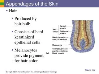
SPECIALIZED CELLS OF THE EPIDERMIS Keratinocytes: • Most common cells of the epidermis. • Provides protection and waterproofing sealant. Melanocytes: • Rounded cells with dendrite-like branches. • Present in Stratum basale. • Produces melanin pigment responsible for the colour of skin. • Melanin is a brown/black pigment that absorbs UV-light.
SPECIALIZED CELLS OF THE EPIDERMIS LangerhansCells (antigen presenting cells): • Non-pigmented granular dendrocytes. • Present in Stratum spinosum. • Nucleus is indented at many places & cytoplasm contains rod-shaped granules. • They participate in immune responses against bacteria and viruses. Merkel Cells: • Found in Stratum basale. • Sensory cells innervated by sensory nerves. • Abundant in fingertips, oral mucosa & hair follicles. • Function as mechanoreceptors.
PIGMENTATION OF SKIN The colour of skin depends upon following factors: • Carotene:yellow-orange pigment (precursor of vitamin A) found in stratum corneum & dermis. • Melanin:produced in epidermis by melanocytes gives black colour to the skin. • Hemoglobin (in blood vessels of dermis): gives pink colour to the skin.
LAYERS OF THE DERMIS • Papillary layer: -Narrow band of loose connective tissue. -In contact with basement membrane of stratum basale. -Dermal papillae (finger- like processes) • Reticular layer: -Dense irregular connective tissue. -Thick elastic fibres. 4 Dermal papilla 3 1 2
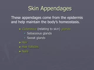
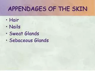
HAIRS: Keratinized filaments derived from invagination of the basal layer of epidermis into the dermis. Parts- a) Root: enclosed by hair follicle. b) Shaft: projects above the surface. Hair follicle: tubular invagination, partly epidermal and partly dermal in origin. APPENDAGES OF THE SKIN
Structure of shaft and root: Medulla Cortex Cuticle Hair follicle: Tubular invagination of epidermis & dermis in which hair root resides. Layers: 3 (inner root sheath, outer root sheath, connective tissue sheath). contd…..
Hair bulb: lower expanded end of hair follicle. Hair papilla: the indentation at the base of hair bulb by part of the dermis. contd…..
ArrectorPilorum Muscle: Smooth muscle innervated by sympathetic nerves. Extends from papillary layer of dermis to the connective tissue sheath of a hair follicle. Contraction of muscle presses the sebaceous gland which squeezes out sebum. Formation of “goose flesh”. contd…..
NAILS: Hardened keratin plates on the dorsal surface of the tips of fingers & toes. Parts: a) Root b) Free border c) Body Nail bed: tissue on which the nail rests. Made up of stratum basale & spinosum. APPENDAGES OF THE SKIN
SEBACEOUS GLANDS: Distributed all over the dermis of the skin, except for the palms & soles. Abundant in the scalp, face, around the apertures of the ear, nose, mouth & anus. APPENDAGES OF THE SKIN
SEBACEOUS GLANDS: Holocrine in nature. Number of alveoli connected to broad duct that opens into hair follicle. Produces an oily secretion called sebum. APPENDAGES OF THE SKIN
SWEATGLANDS (SUDORIFEROUS GLANDS) • Absent from lips, glans & nail bed. • Mode of secretion: merocrine • Simple tubular gland • 2 parts: (a) Secretory portion (b) Excretory duct Secretory portion: • Twisted coil like structure with simple cuboidal epithelium. • 3 types of cells: clear cells, dark cells, myoepithelial cells. Excretory duct: • Long & extends from secretory portion to surface of epidermis.
contd…. 2 types: Eccrine: • Most numerous in the soles & palms. • Produces thin watery secretion. Apocrine: • Confined to axilla, eyelids (Moll’s glands), nipple & areola of breast, perianal region, and the external genitalia. • Produces thick odourous secretion. • Ceruminous glands & lactating mammary glands are modified apocrine sweat glands.
Langer’s or Cleavage lines • The lines along which the fibre bundles run. • Represent the natural lines along which the skin tends to split when penetrated. • Incisions in the direction of these lines gape much less than those at right angles to them.
Linea gravidarum • Rupture of fibre bundles of dermis due to excessive stretching result in prominent white lines. • Seen in anterior abdominal wall in pregnancy.
Rule of Nine: To estimate the extent of damaged skin in burn injuries. • First degree burn- only epidermis. • Second degree burn- epidermis + upper region of dermis. • Third degree burn- entire thickness of skin.
Dermatomes • The strip of skin supplied by a single spinal nerve is called dermatome.
References 1. diFiore’s Atlas of Histology with functional Correlations, 12th Edition. 2. Textbook of Human Histology. Inderbir Singh, 1st Edition. 3. Textbook of Histology. GP Pal, 3rd Edition.
MCQ Q1. Which layer is present only in thick skin: • Stratum basale • Stratum spinosum • Stratum granulosum • Stratum lucidum
MCQ Q2. The characteristic feature of reticular layer of dermis is: • High mitotic activity • Contains keratin granules • Dense irregular connective tissue • Finger like processes
MCQ Q3. Secretion of sebaceous glands is aided by contraction of: • Arrectorpilorum muscle • Myoepithelial cells • Papillary layer of dermis • Reticular layer of dermis
MCQ Q4. Langerhans cells are present in: • Stratum basale • Stratum spinosum • Stratum granulosum • Stratum lucidum
MCQ Q5. The sensory cells of epidermis are: • Melanocytes • Keratinocytes • Langerhans cells • Merkel cells