1 / 64

Retinal Detachment

Retinal Detachment. Dr Soujanya K Assistant Professor, YMCH Mangalore. Specific learning objectives. To understand the definition of RD To describe pathogenesis of different types of RD To describe the principle of management of RD. Retinal detachment - definition.

price
Download Presentation

Retinal Detachment

An Image/Link below is provided (as is) to download presentation Download Policy: Content on the Website is provided to you AS IS for your information and personal use and may not be sold / licensed / shared on other websites without getting consent from its author. Content is provided to you AS IS for your information and personal use only. Download presentation by click this link. While downloading, if for some reason you are not able to download a presentation, the publisher may have deleted the file from their server. During download, if you can't get a presentation, the file might be deleted by the publisher.

E N D

Presentation Transcript


  1. Retinal Detachment Dr Soujanya K Assistant Professor, YMCH Mangalore

  2. Specific learning objectives • To understand the definition of RD • To describe pathogenesis of different types of RD • To describe the principle of management of RD

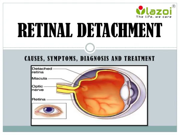
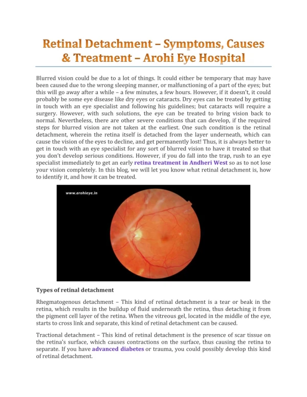
  3. Retinal detachment - definition • Separation of the neurosensory retina (NSR) from the retinal pigment epithelium (RPE).

  4. Embryology

  5. Potential space Embryology – Neurosensory retina and RPE

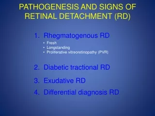
  6. Types of RD 1. Rhegmatogenous RD - Primary 2. Tractional RD 3. Exudative RD Secondary

  7. Rhegmatogenous RD

  8. Vitreous is a gel-like substance in the eye

  9. Posterior Vitreous detachment

  10. Posterior Vitreous detachment

  11. Retinal tear

  12. Vitreous liquifaction

  13. Vitreous liquifaction

  14. Seepage of liquid vitreous through the hole

  15. Retinal Detachment

  16. Exudative RD

  17. Retina Being Pushed Away

  18. Choroidal haemorrhage • Exudativechoroiditis/retinopathy • Angiomatosis • Toxaemia of pregnancy By fluid

  19. By tumor

  20. Retina Being Pushed Away by tumor

  21. Retina Being Pushed Away by tumor

  22. Tractional RD

  23. Proliferative retinopathy Retinopathy of prematurity.

  24. contraction of fibrous tissue in the vitreous

  25. Retina is mechanically pulled away from its bed

  26. Tractional RD

  27. Tractional RD

  28. Rhegmatogenous Retinal Detachment Risk factors

  29. Myopia. • Aphakia • Retinal degenerations • Lattice degeneration • Snail track degeneration • White-with-pressure and • white-without-or pressure • Acquired retinoschisis • Focal pigment clumps • Trauma • PVD

  30. Lattice degenration

  31. Snail track degeneration

  32. White Without Pressure

  33. Acquired retinoschisis

  34. symptoms

  35. Signs • IOP may be low • Marcus Gunn pupil/ RAPD • Altered red reflex

  36. Slightly opaque and Corrugated retinal blood vessels appear darker convex configuration

  37. Treatment

  38. 3 Principles • Sealing the break • Draining sub-retinal fluid • Maintaining chorioretinalaposition

More Related