Download
1 / 45
670 likes | 1.91k Views
Skin & Wound Infections. Dr. Ihsan Edan Alsaimary Assistant Professor Department Of Microbiology College Of Medicine University Of Basrah. Normal function of skin: prevent colonization and invasion of underlying tissue by potential microbial pathogens

E N D
Skin & Wound Infections Dr. Ihsan Edan Alsaimary Assistant Professor Department Of Microbiology College Of Medicine University Of Basrah
Normal function of skin: prevent colonization and invasion of underlying tissue by potential microbial pathogens • Loss of skin integrity (wound) provides moist and nutritious environment for microbial proliferation • Presence of foreign material and necrotic tissue facilitates microbial proliferation (“dirty” wound)
SKIN AND WOUND INFECTIONS Entry Skin (pores, hair follicles) Wounds (scratches, cuts, burns) Insect & animal bites Diseases Localized infections with local and/or systemic effect Systemic infections Multiplication Extracellular Intracellular (??) Damage Toxins, exoenzymes Host immune response
DEFENSES • Dry : usual infection sites are wet areas, skin folds, armpit, groin • Acidic (pH 5.0) • Temperature less than 37oC: Some pathogens grow best <37oC • Lysozyme and toxic lipids: pore, hair follicles, sweat gland • Resident microflora : mainly Gr+ • Skin-associated lymphoid tissue (SALT)
COMMON SKIN INFECTIONS Local infections • Impetigo: epidermis • Folliculitis: infection of hair follicles • Furuncles (boil): extension of folliculitis • Carbuncles: furuncles coalesce and extend to the deeper subcutaneous tissue fever and chills • Skin abscesses : • Localized infection of dermis and subcutaneous tissue • Erysipelas - dermal lymphatics • Cellulitis - subcutaneous fat layer Systemic effect • Toxic shock • Sepsis
Cellulitis • Acute, spreading infectious process affecting epidermis and dermis • Inflammation with little or no necrosis, edema • Lymphatic involvement • Fever, chills, leukocytosis • Bacteremia up to 30% of cases • Complications: • Abscess and osteomyelitis • S. aureus • Streptococcus pyogenes (group A streptococcus)
Staphylococcus aureus Local skin infections: Impetigo, Folliculitis, Furuncles (boil), Carbuncles Systemic infections: Staphylococcal Scalded skin syndrom (SSSS) • Toxic shock syndrom controlAfter 12h incubation with S. aureus TSST-1 Sepsis Endocarditis Osteomyelitis
Pathogenesis • Encounter : Broken skin or mucous membrane – humans only • Spread : can spread hematogenously ANYWHERE to cause abscesses or osteomyelitis or septic arthritis • can also cause toxigenic infections --- toxin spreads • Multiplication : grows well anywhere • Avoid Host Immune Response : coagulase, catalase, leukotoxins, Fc binding protein, adhesins • Damage : inflammation, superantigens, degradative enzymes • Transmission : contact from fomites or direct person-person contact Major defenses against S. aureus • C3b • Activated by Staph cell wall fragments • Opsonizes the bacteria • Enhances phagocytosis • Chemotaxis – attracts neutrophils • Neutrophils – engulf the bacteria • Intracellular killing by O2 radicals Conditions predispose to Staph infections • C3 hypercatabolism • Neutropenia • Chemotherapy • Cyclic neutropenia • Chronic granulomatous disease • reduced production of H2O2 and O2 • “Lazy leukocyte” (chemotaxis deficiency) Virulence Factors 1- Fibronectin binding protein 2- Toxins • Superantigen (TSST-1) • Dermonecrotic toxin • Enterotoxin 3- Enzymes • Hyaluronidase, collagenase • Lipase • Coagulase • Hemolysin, leukocidin • Nuclease, protease 4- Fc binding protein
Staphylococcal scalded skin syndrom(SSSS) • Dermonecrotic toxin (exfoliative toxin) • Bullous exfoliative dermatitis Toxic shock syndrome • Toxic shock syndrome toxin(TSST-1) • Super antigen • Produced by 5-25% isolates • Tampon or infected wound : • Fever • Rash • Exfoliation of skin • Shock (death rate 3%) TSST-1 Structure : 22 KD secreted protein Mechanism of action • Interacts with MHC-II and T-cell receptors • Stimulate the release of cytokines (IL-1, IL-2 and TNF) Treatments • Synthetic antibodies and peptides: Block interaction with MHC-II, preventing T cell activation • Immunosuppressants : Prevent T-cell activation and cytokine releases • Corticosteroids : Reduce inflammatory effects
Streptococcus pyogenes Local infections • Impetigo • Erysipelas • Cellulitis • Necrotizing fasciitis (flesh-eating bacterium) Systemic effect • Streptococcal toxic shock-like syndrome (STSS) • Spe (similar to TSS by S. aureus) • Scarlet fever (pyrogenic toxin by lysogenized φ) • Post-infection • Rheumatic fever (associated with pharyngitis) • Glomerulonephritis Virulence factors 1- Adhesins • M protein (fibrillar Ag) • Fibronectin binding proteins (Protein F) • Lipoteichoic acid (LTA) 2- Hyaluronic acid capsule 3-Invasins • Streptolysins (S & O) • Hyaluronidase • Streptokinases : activates blood clot dissolving protein-plasminogen (human specific) • Dnase 4-Exotoxins Pyrogenic (erythrogenic) toxin - Spe Scarlet fever Toxic shock syndrome
Clostridial infections (soil) Anaerobic Gr+, spore-forming 1- Wound botulism (Clostridium botulinum) Deep wounds: • anaerobic environment • Botulinum toxin (BT) leaks into blood BT enters neurons • Inhibits acetylcholine release in neuromuscular junction • prevent muscle contraction : flaccid paralysis Therapy • Antibiotics • Antitoxin • Supportive therapy Botulinum toxin Structure :A & B Chains (50/100 KD) by disulfide bond Mechanism of action Protease • Cleave fusion protein (SNAP-25) at neuromuscular junction • Prevent vesicles from anchoring to the membrane • Inhibit acetylcholine release Application in the cosmetics :Botox and Dysport Distinguish “food botulinum” :Premade toxin (canned beans) “infant botulinum” :Spore in foods (honey)
2- Tetanus (Clostridium tetani) Local infection with systemic effect Early symptoms • Locked jaw • Stiffness in the neck and abdomen • Difficulty swallowing Late symptoms • Fever • Elevated blood pressure • Severe muscle spasm Tetanospasmin (A & B toxin) Released when cell lyses A domain – a zinc endopeptidase, attacks the vesicleassociated membrane protein (VAMP), degrading synaptobrevin, thus stop the release of inhibitory neurotransmitters GABA and glycine B domain – binds disialogangliosides (GD2 and GD1b) on the neuron membrane Acts in spinal cord, prevents the inhibition of contraction of opposing sets ofmuscles – hence, tetanic or spastic paralysis. Vaccine • Tetanus toxoid • DTaP for <7 yr, Td for >7 yr
3- Gas gangrene (Clostridium perfringens) Alpha toxin (phospholipase C): Zinc metallophospholipase hemolysis and bleeding Gas formation Mechanism (by host cell) Content: N2 (74.5%), O2 (16.1%), CO2 (3.4%), H2 (5.9%) Myonecrosis, shock, renal failure and death Alpha toxin Structure : Single 40KD protein Mechanism of action 1- C-terminal domain binds phospholipid bilayer 2- N-terminal domain has phospholipase activity 3- The hydrolysis of phosphatidyl choline produces diacylglycerol which activates a variety of second messenger pathways. The end result includes edema due to increased vascular permeability. Treatment • Debridement and excision • Antibiotics (prevent further spreading) • Hyperbaric oxygen therapy : Inhibit or kill the anaerobic bacteria
Cutaneous Anthrax Bacilllus anthracis Gr+ and spore forming Farm animals are major reservoir Inhalation, GI, cutaneous Virulence factors: Capsules Edema factor (A) + Protective antigen (B) Lethal factor (A) + Protective antigen (B) Vaccine Toxoid (protective antigen) Effective in short term but not long term
Other Skin and Mucus Membrane Infections Staphylococcus epidermidis Catheters and prostheses Vibrio vulnificus From shellfish and salt water Obligate anaerobes Puncture wounds Deep wounds Impaired blood supply Gram negative bacteria Decubitus ulcer (bed sores) After intestinal “spill” Pseudomonas aeruginosa Catheters and prostheses Burns Surgical wounds
Fungal infection (dermatophytes) • Tinea(pedis, corporis, cruris, manuum, facei, capitis, unguium) • Id reaction : allergic dermatophytids • Dermatoplytosiscomplex : mixed fungal and bacterial infection of toe web
Laboratory diagnosis –Dermatophyte infection • Skin scraping –choose the active margin, collect scale with colourpaper, (dry up the oily or wet skin with alcohol or ether in the old days before scraping), can send to laboratory by post • Hair plugging (NOT cutting) –choose the short broken hair (mostly likely found in the margin of bald patch), can be guided by Wood’s light; sample with specific brush; sample the pets • Nail clipping –scrape the sub ungual hyperkeratosis as proximal as possible, maximum amount of disease nail material, may use a small punch biopsy needle to sample in proximal subungualonychomycosis, scraping of the nail surface in superficial white onychomycosis • Wet mount –dissolve the keratin material with KOH, direct examination with or without stain (e.g. Parker stain) [Sn12% & Sp 93%] • Culture with Sabouraudagar (if mould [non-dermatophytefilamentous fungi] infection is suspected, culture without cycloheximideis required) • Nail clipping for histology (with special stain) can be performed in those persistent culture negative cases (with clinical features of fungal infection)
Fungal infection (yeast & Pityrosporum/Malassezia) • Candidiasis: intertriginousarea, napkin area, chronic paronychia, chronic mucocutaneouscandidiasis, balanitis, oral thrush • Pityrosporum: pityriasisversicolor, pityrosporumfolliculitis(seborrhoeicdermatitis, pityriasiscapitis) • Trichosporosis: pied
Viral skin infections • HSV: cold sore, genital herpes • VZV: Chicken pox,zoster • HPV: wart • Pox virus : molluscumcontagiosum • Viral exanthemata :measle, rubella,roseola infantum, erythemainfectiosum, • Others: Hand foot mouth disease; (EBV, HIV)
Parasitic infestation • Mite: Sarcoptes scabei • Lice: head lice, body lice, pubic lice • Others: cutaneous larva migran, amoeba, myiasis, etc
wound infections • Pathophysiology • Acute wounds: external damage to intact skin (surgical wounds, bites, burns, gunshots, minor cuts and abrasions) • Chronic wounds: endogenous mechanisms compromising epidermal and dermal tissue (impaired arterial supply or venous drainage, diabetes mellitus, poor nutrition, immunosuppression, sustained external skin pressure)
Pathophysiology of wound infection • Microbial colonization precedes wound infection • If tissue devitalized and/or host immunity compromised, conditions optimal for microbial growth and invasion follows colonization • Source of microorganisms: exogenous (environmental), surrounding skin, and endogenous (mucous membranes of gastrointestinal tract and genitourinary tract, oropharyngeal cavity)
Predisposing factors for wound infection • Poor blood perfusion with hypoxia (pO2 < 20 mm Hg) inhibits granulation tissue response and wound repair • Cell death and tissue necrosis due to hypoxia creates ideal growth conditions for wound microflora • Hypoxia compromises oxygen radical dependent killing of bacteria by polymorphonuclear neutrophils
Pathophysiology of wound infection • Density of microorganisms the critical factor determining whether or not a wound heals • Presence of specific microbial pathogens of primary significance in delayed wound healing • Most likely both factors important in delayed wound healing due to infection
Pathophysiology of wound infection • Healing of decubitus ulcers occurs only when bacterial load <106 cfu/ml of wound fluid • Acute and chronic wound infection occurs with microbial load >104 (complex extremity wounds) or >105 cfu/g of wound tissue • Single microorganism on Gram’s stain reliably predicts >105 cfu’s/g of wound tissue • Presence of bacterial cells on Gram’s stain of burn wounds consistently correlates with >106 organisms per swab specimen • Critical microbial load for wound infection appears to be 104-106cfu/g wound tissue or ml wound fluid, and 106cfu/wound swab specimen
Microbiology of wound infection: the Big Three1 • Streptococcus pyogenes (capable of wound infection <105 cfu/g wound tissue)2 • Staphylococcus aureus • Pseudomonas aeruginosa 1Associated with monomicrobial and polymicrobial wound infection 2And other pyogenic β-hemolytic streptococci
Microbiology of wound infection: Obligate Anaerobic Bacteria1 • Bacteroides • Porphyromonas (pigmented) • Prevotella (pigmented and non-pigmented) • Fusobacterium • Peptostreptococcus • Clostridium2 1Primarily associated with polymicrobial aerobic and anaerobic bacterial wound infection 2Monomicrobial infection by Clostridiumperfringes in myonecrosis (gas gangrene) (distinctive Gram’s stain showing large “boxcar shaped” gram-positive rods with a paucity of inflammatory leukocytes
Polymicrobial wound infection: mechanisms • Oxygen consumption by aerobic bacteria induces tissue hypoxia and favorable growth conditions for anerobic bacteria • Nutrients produced by one organism supports growth of other fastidious and potentially pathogenic organisms
Polymicrobial wound infection: mechanisms • Vitamin K production by Staphylococcus aureus supports growth of vitamin K-dependent Prevotella melaninogenica • Succinate produced by Klebsiella pneumoniae a critical growth factor for Prevotella melaninogenica
Microbiology of wound infection: surgical wounds1,2 • Staphylococcus aureus (191 patients, 28.2%) • Pseudomonas aeruginosa (170 patients, 25.2%) • Escherichia coli (53 patients, 7.8%) • Staphylococcus epidermidis (48 patients, 7.1%) • Enterococcus faecalis (38 patients, 5.6%) • Anaerobic bacteria (21 patients) 1n=672 surgery patients with wound infections 2Giacometti et al., JCM 38:918-922 (2000)
Microbiology of wound infection: surgical wounds1 • Superficial wounds, surgical incisions: streptococci, staphylococci • Deep wounds: GI, female genital tract, and oropharyngeal-streptococci, staphylococci, gram-negative enterics, enterococci, Bacteroides, other anaerobes; other-streptococci, staphylococci, gram-negative enterics • Gangrenous 24-48 hr after surgery: group A streptococci, clostridia • Necrotizing >4 days after surgery: polymicrobial (aerobic and anaerobic) 1Nichols and Florman, CID 33(Suppl 2):S84-93 (2001)
Microbiology of wound infection: diabetic foot infections1 • Cellulitis: β-hemolytic streptococci (A, B, C, G), Staphylococcus aureus • Infected ulcer (no antibiotic treatment): same as cellulitis, often monomicrobial • Infected ulcer that is chronic or with previous antibiotic: S. aureus, β-hemolytic streptococci, Enterobacteriaceae (usually polymicrobial) • Macerated ulcer due to soaking: Pseudomonas aeruginosa (usually polymicrobial) • Long-duration non-healing ulcers with prolonged, broad-spectrum antibiotic treatment: S. aureus (MRSA), coagulase-negative staphylococci, enterococci (VRE), diphtheroids, Enterobacteriaceae (ESBL resistance), Pseudomonas, nonfermentative gram-negative’s, possibly fungi (usually polymicrobial) • “Fetid foot” with extensive necrosis, gangrene, and malodorous: Mixed aerobic gram-positive cocci, Enterobacteriace, nonfermentative gram-negative’s, obligate anaerobes 1Lipsky et al., CID 39:885-910 (2004)
Microbiology of wound infection: burn wound infections1 • Staphylococcus aureus (22.9%) • Pseudomonas aeruginosa (20.9%) • Pseudomonas species(7.2%) • Escherichia coli (6.7%) • Group D Streptococcus (5.0%) • Enterococcus faecalis (4.2%) 1Bacteria constituting >4% of organisms that were recovered from 1,267 burn wound infections during 1974-1978 (CDC, Mayhall, CID 37:543-550 (2003)
Microbiology of wound infection: burn wound infections1 • Staphylococcus aureus (23.0%) • Pseudomonas aeruginosa (19.3%) • Enterococcus species (11.0%) • Enterobacter species (9.6%) • Escherichia coli (7.2%) • Coagulase-negative Staphylococcus (4.3%) 1Bacteria constituting >4% of organisms that were recovered from 1,234 burn wound infections during 1980-1998 (CDC, Mayhall, CID 37:543-550 (2003)
Microbiology of wound infection: human bite infections1,2 • Streptococcus (84%) • Staphylococcus (54%) • Prevotella (36%) • Fusobacterium (34%) • Eikenella corrodens (30%) 1Bacteria recovered from >30% of 50 patients with infected human bite injuries. 2Talan et al., CID 37:1481-1489 (2003)
Microbiology of wound infection: animal bite infections1,2 Same as human bites with the addition of: • Pasteurella multocida • Neisseria weaveri • Staphylococcus intermedius 1Goldstein, Mandell, Douglas, and Bennett’s Principles and Practice of ID, pp. 3552-3556 (2005) 2Capitini et al., CID 34:e74-74 (2002).
Clinical signs of wound infection • Purulent discharge • Painful spreading erythema • Failure to heal
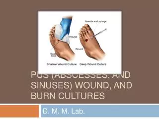
Wound Specimens • Tissue • Wound fluid (purulent exudate) • Superficial swabs • Basic principle of specimen collection: Only wounds with clinical signs of infection, are deteriorating, or fail to heal should be sampled for Gram’s stain and culture
Gram’s stain of wound specimens • Presence of bacteria by Gram’s stain indicates 105 to 106 organisms/g wound tissue or ml wound fluid • Types of organisms present on Gram’s stain should be correlated with culture results to recognize predominant organisms that don’t grow aerobically
Culture of wound specimens • Facultative anaerobic and aerobic bacteria of primary importance in wound infection • Media for aerobic culture of wound specimens include sheep blood, chocolate, and MacConkey agar • Media for anaerobic culture of wound specimens include brucella blood agar, laked blood with kanamycin and vancomycin, Bacteroides bile esculin, and anaerobic colistin-naladixic acid
Culture of wound specimens • Correlate growth of facultative anaerobic and aerobic bacteria with gram-stain morphotypes • If Staphylococcus aureus, β-hemolytic Streptococcus, and/or Pseudomonas aeruginosa present in any numbers, identify with susceptibility testing • If coagulase-negative Staphylococcus, Corynebacterium, and/or Enterococcus present in moderate to many numbers, and growth explains gram-stain results, report as genus and full identification/susceptibility available by request. • If Enterobacteriaceae, or non-fermenters other than Pseudomonas aeruginosa present in moderate to many numbers, and growth explains gram-stain results, identify with susceptibility testing • If >4 facultatively anaerobic or aerobic bacteria detected in culture by these criteria, obtain a technical consult • Report obligate anaerboic bacteria as polymicrobial flora
Culture of wound specimens • If facultative anaerobic and aerobic bacteria recovered in culture do not correlate with one or more gram-stain morphotypes, review and repeat the Gram’s stain • If aerobic cultures still do not explain Gram’s stain upon review and repeat, examine anaerobic cultures • If obligate anaerobic bacteria in moderate to many numbers correlate with gram-stain morphotype(s) not explained by aerobic cultures, identify by genus and report susceptibility available by physician request • If > 4 organisms detected in culture by these criteria, obtain a technical consult
Genus identification of anaerobic bacteria in wound culture • Bacterioides: growth in 20% bile • Porphyromonas: vancomycin susceptibile, kanamycin resistant, colistin resistant1 • Prevotella: vancomycin resistant, kanamycin resistant, colistin susceptibile1 • Fusobacterium: vancomycin resistant, kanamycin susceptible, colistin susceptibile 1+/- pigmentation on laked blood
Genus identification of anaerobic bacteria in wound culture • Clostridium: large gram-positive rods with sporulation • Peptostreptococcus: kanamycin resistant and sodium polyanethol sulfonate susceptible, or kanamycin susceptibile and sodium polyanethol sulfonate resistant • Perform aerotolerance on all anaerobic isolates to be reported
Wound specimen negative by direct Gram’s stain • Review and repeat Gram’s stain. If confirmed negative, proceed as outlined below • Identification and susceptibility testing of Staphylococcus aureus, β-hemolytic Streptococcus, and Pseudomonas aeruginosa only • Other culture isolates reported as “polymicrobial flora”