Download
1 / 33
330 likes | 792 Views
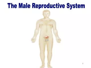
The Male Reproductive System. Differentiation of the Embryo. Gonad Development. The male and Female gonads are derived, embryologically, from the same site in the body.

E N D
Gonad Development • The male and Female gonads are derived, embryologically, from the same site in the body. • All fetuses start out as female. Under the influence of the Y chromosome “masculization” occurs. Default development is female. A gene on the Y chromosome identified as SRY starts a chain of gene-expression reactions that causes STEROIDSYNTHESIS, specifically TESTOSTERONE which causes the testis development pathway. Without testosterone, the “default” or female pathway is followed.
Sexual Dimorphisms • The glands penis and its prepuce and the clitoris and its prepuce are actually sexually dimorphic analogous structures. • They both contain a shaft, glands and spongy tissue which becomes engorged with blood when stimulated. They are highly sensitive.
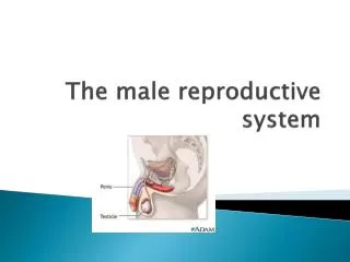
Penile Components • The scrotum is the pouch that contains the testes and maintains their temperature at approximately 93.6 degree Fahrenheit to allow for spermatogenesis. • Epididymis where the mature sperm are stored(located at surface of each testis). • The Vas Deferentia, which join the ducts of the seminal vesicles, provide secretions that nurture the sperm. • Cowper’s Glands flank the urethra and secrete clear, mucus-like fluid that appears as preejaculatory fluid.
Human Male Anatomy • Sperm are produced in the testes which are located in the scrotum • Before birth the testes form in the embryo’s abdomen and then descend into the scrotum • Sperm require a temp. that is 3 degrees Celsius below body temp. That is why they are outside the abdomen. Muscles help regulate temp.
Components • Scrotum • Testes • Duct system • Penis • Accessory glands 24.1
Scrotum • Skin • Dartos muscle (smooth) • Septum • Cremaster muscle (skeletal) 24.2
Scrotum • Temperature • Optimal for sperm development is 3°C below body temp (~91 F) • Controlled by muscles • Spermatic cord • Testicular artery • Plexus of veins • Nerves • vas deferens 24.2
Testes – gross anatomy • ~4 cm long, 2.5 cm wide • Tunica vaginalis • Tunica albuginea (capsule) • Septa • 250-300 lobules • Seminiferous tubules • Epididymis 24.3
Descent of the Testes 3 mos. 7 mos. birth 24.29
Seminiferous tubules • Spermatogenic cells • Spermatogenesis begins at puberty • ~400 million sperm/day 24.3b
Spermatogenic cells • Spermatogonia • On basal lamina • Mitosis • Primary spermatocytes • 1st meiotic division • Secondary spermatocytes • 2nd meiotic division • Spermatid • Haploid • 4 from each 1° spermatocyte 24.4b
Seminiferous tubules • Spermatogenesis • Spermatogonia • 1° spermatocyte • 2° spermatocyte • Spermatid 24.4c
Hormones • Spermatogenesis controlled by • FSH (follicle stimulating hormone) from pituitary • Testosterone from Leydig (interstitial) cells of testes • secretions from Sertoli cells
Spermiogenesis • Spermatid spermatozoan • Head • Nucleus • Acrosome (lysosome) • Midpiece • Base of tail • Mitochondria • Tail • Long flagellum 24.5
Spermiogenesis • Spermatid spermatozoan 24.5
Sustentacular (Sertoli) cells • Nurse cells • Extend from basal lamina to lumen • Connected to each other by tight junctions • Nourish spermatogenic cells • Transport spermatogenic cells • Phagocytize excess cytoplasm • Secretions regulate spermatogenesis 24.4c
Sustentacular cells 2 Sertoli cells with associated spermatogenesis cells. 24.4c
Interstitial (Leydig) cells • Secretes testosterone • sER for steroid production • Controlled by LH from pituitary 24.3b
Ducts • Epididymis • Ductus deferens • Ejaculatory duct • (urethra) 24.1
Epididymis • Located in scrotum • Between seminiferous tubules and vas deferens • ~6 m long 24.3a
Ductus (vas) deferens • From scrotum to pelvis • Forms ejaculatory duct with seminal vesicle duct • Empties into prostatic urethra • Thick muscularis • Propels sperm during ejaculation 24.1
Urethra • Shared with urinary system • Prostatic urethra • Membranous urethra • Spongy (penile) urethra 24.8
Accessory Glands • Seminal vesicle • 60% of semen • Fructose to nourish sperm 24.1
Prostate Gland • Prostate gland • Surrounds prostatic urethra • 30% of semen • Supports sperm 24.9
Penis • Thin skin • Spongy urethra • Erectile bodies • Corpus spongiosum • 2 corpora cavernosa • Capsule • Vascular spaces 24.8
Epididymis slide . • In PowerPoint file for male reproductive system. • http://www.southalabama.edu/biomedical/311Anatomy/BMD311.htm
Erection • Sexual excitement (parasympathetic) • blood flow to erectile bodies • Squeezes veins shut • blood pressure in erectile bodies 24.8
Ejaculation • Sympathetic activation • Peristaltic contraction of smooth muscles in ducts and glands - ejaculation • Constriction of arteries • blood pressure in erectile bodies 24.8